
Inquiry Basket
 Loading ......
Mycoplasma pneumoniae (MP) is a small genome microorganism. It is the smallest free-living, cell-wall-less, self-replicating prokaryotic organism. MP is a major cause of acute respiratory tract infections and pneumonia in humans. Its infection is usually self-limiting, but in some patients, MP infection can cause severe upper and lower respiratory symptoms and can be life-threatening, especially in children and adolescents. Community-acquired pneumonia (CAP) has a high morbidity and mortality rate in children and is a major threat to global public health. Mycoplasma pneumoniae pneumonia (MPP) is one of the most common types of CAP. Some studies suggest that it accounts for more than 50% of CAP in children over 5 years of age. Studies have shown that the MP infection rate in children with acute asthma can be as high as 46%. In addition to invading the respiratory tract and causing laryngitis, pharyngitis, bronchitis, atypical pneumonia and exacerbated asthma, MP infection can also cause extrapulmonary complications through complex immune mechanisms involving the skin, brain, kidney, musculoskeletal, digestive and other systems, leading to some rare clinical symptoms. MP mainly adheres to the host respiratory epithelial cells through adhesion organelles, thereby destroying the integrity of the airway mucosa and the immune defence function, and then causing inflammatory responses. Some studies have shown that MP mutants that have lost their ability to adhere are not pathogenic. As one of the major adhesion proteins, the P1 protein of MP not only plays an important role in the adhesion process between MP and host cells, but also participates in the gliding of MP on the airway surface. This gliding movement allows MP to move from the ciliary tip of bronchial ciliated epithelial cells to the cell surface, releasing toxins, hydrogen peroxide and superoxide free radicals, leading to cell damage. Adhesion and motility of MP are prerequisites for its colonisation of the host airways. This article reviews the gene structure, protein function and pathogenic mechanism of the P1 protein to elucidate the mechanism of action of the P1 protein in MP adhesion, gliding and pathogenicity.
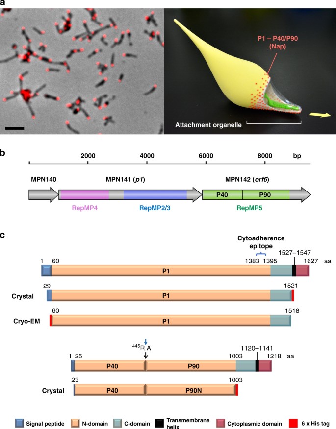 Figure 1. The Nap complex in M. pneumoniae. (Sources: Vizarraga D, et al. 2020)
At the initial stage of infection, MP recognises host sialylated and sulphated oligosaccharide receptors, adheres to the host bronchial epithelium via terminal adhesion structures and induces metabolic and ultrastructural changes in infected cells. The adhesion organelle at the tip of MP comprises a structure containing three types of proteins: adhesion proteins, chaperone proteins and helper proteins. Adhesion proteins are mainly P1 proteins and P30 proteins, and helper proteins include P40 proteins, P90 proteins, P65 proteins, P116 proteins and high molecular weight (HMW) proteins 1-5. The adhesion organelle consists of two parts: the surface and the inner structure. Its core is composed of a "dumbbell-button-bowl" complex, which includes a terminal button composed of HMW2, HMW3 and P65 proteins, a paired plate composed of HMW1, HMW2, CpsG and HMW3, and a bowl-shaped complex composed of Lon, P24, Topj, P200, P41, MPN387 and HMW2. Interaction between this adhesion organelle and the host airway epithelium can induce cytoskeletal rearrangements in host cells, thereby promoting signal transduction after infection. Based on its adhesion, MP slides on the host airway surface and after invading the body tissue, it slides forward along the direction of the adhesion organelle at a speed of about 1 μm/s.
The total length of the MP genome is 823kb and the P1 gene mgpA is 4884bp with a GC content of 53.3%. MP colonises the respiratory tract with a unique adhesion organelle. The P1 protein is an essential molecule for MP to successfully colonise and cause disease in the host respiratory epithelium. The molecular weight of the P1 protein is 170 kDa, making it the largest adhesion protein of MP and one of the major proteins that make up its adhesion organelle. The researchers identified a key sequence of the P1 protein through experiments and found that the proline-rich C-terminus of the P1 protein may be the main factor involved in its terminal attachment to the mucosa and plays an important role in the formation and function of adhesion organelles. The correct positioning of P1 in the adhesion organelle is a prerequisite for MP to adhere to the host cell. After MP contacts the target host cell, the P1 protein dispersed on its surface is translocated to the surface of the adhesion organelle at the top of MP. Studies have shown that the P1 protein can directly interact with specific receptor structures on the host cell membrane, is involved in regulating recognition between MP and host respiratory epithelial cells, and plays an important role in the gliding movement of MP.
P1 protein is an important surface structural protein for MP to adhere to organelles. MP promotes its sliding on the surface of host respiratory epithelial cells through the adhesion of P1 protein and the deformation movement of the "dumbbell-button-bowl" complex in which it participates. MP relies on the binding and separation of P1 protein and host cell surface receptors (i.e., sialic acid oligosaccharides) to enable pathogens to continuously slide forward. Studies have shown that P1 protein can directly bind to sialic acid residues of glycoproteins on the surface of host airway epithelial cells. As a receptor for P1 protein to adhere to host cells, sialylated oligosaccharides provide support for MP adhesion, colonization and sliding movement in the airway, which is an important reason for P1 protein to participate in MP pathogenicity.
The interaction between P1 protein and sialic acid is affected by factors such as sialic acid density, type and microenvironment. Studies have found that P1 protein can bind to α-2,3 and α-2,6 sialic acid lactose in a concentration-dependent manner and support P1 protein adhesion. The specific connection between the P1 protein complex and the two sialic acids may lead to different pathogen presentation patterns, and the relative abundance of the two sialic acids may have a profound impact on the colonization and gliding movement of MP in the airway. Studies have shown that MP exhibits a higher binding affinity for α-2,3-linked sialic acids, and the gliding frequency increases accordingly with the increase in the density of α-2,3-sialyllactose. When the concentration of α-2,3-sialyllactose is greater than 8%, the ability of P1 protein to bind to it and glide is the strongest. In addition, the number of P1 adhesin complexes bound to α-2,3-sialyllactose also promotes cell sliding to a certain extent.
After MP infection, P1 protein directly contacts sialic acid residues to induce inflammatory response, and sialidase (neuraminidase) can reduce the induction of MP on inflammation. In the MP-induced mast cell or factor response, P1 protein directly contacts sialic acid residues on the surface of mast cells, mast cells are activated, and inflammatory damage is caused.
References
| Target | Cat. No. | Product Name | Host | Isotype | Application | |
| M. pneumoniae P1 | CABT-B8818 | Anti-M. pneumoniae P1 monoclonal antibody, clone C2059N | Mouse | IgG1 | ELISA(Cap), IF | Inquiry |
| CABT-B8819 | Anti-M. pneumoniae P1 monoclonal antibody, clone C2054N | Mouse | IgG2b | ELISA(Det), IF | Inquiry |
| Target | Cat. No. | Product Name | Expression System | Tag/Conjugate | Application | |
| M. pneumoniae P1 | DAGC083 | Native Mycoplasma Pneumoniae P1 Antigen | N/A | Unconjugated | Inquiry | |
| DAG-WT2194 | Recombinant M. pneumoniae Adhesin P1&P30 complex Protein [His] | E. coli | His | Immunoassays | Inquiry |
Loading ......
