
Inquiry Basket
 Loading ......
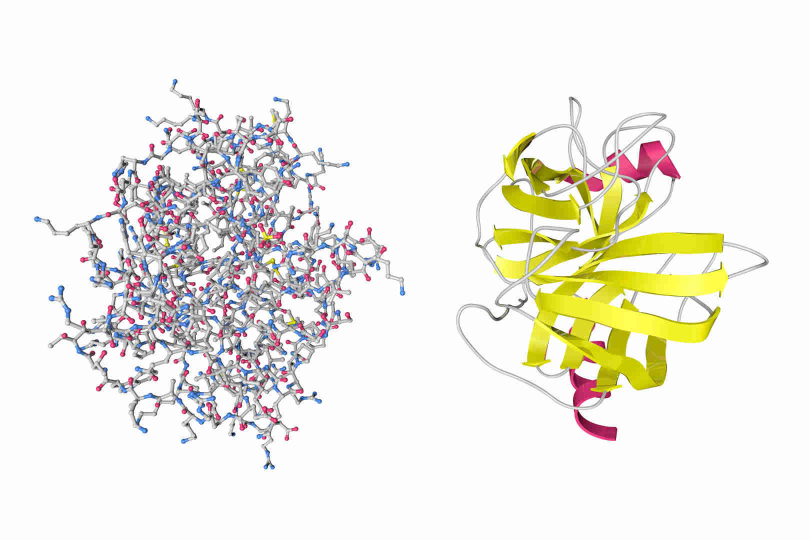
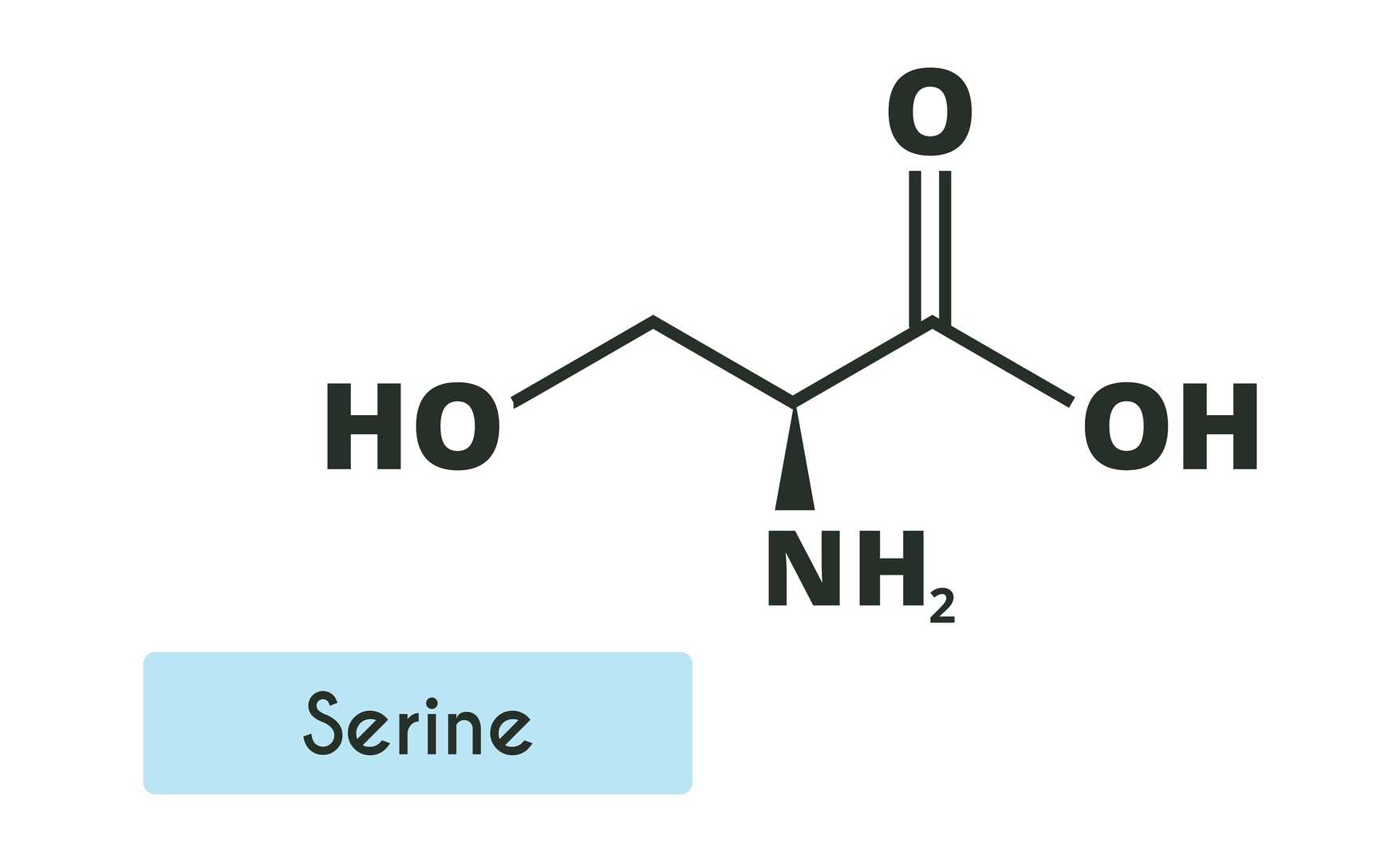
Serine proteases are a group of proteases which are characterised by the presence of a serine (SER) residue in the active site that participates directly in catalysis of substrate proteins by nucleophilic attack on the peptide bond. They are the most studied and largest group of proteases, making up more than one third of all proteases. Serine proteases are found from bacteria to humans and are involved in a broad spectrum of essential biological functions including metabolism, immune and inflammatory responses, as well as signal transduction.
Serine is coded for by six codons (UCU, UCC, UCA, UCG, AGU and AGC), more than any other amino acid. Serine can be produced by de novo synthesis or from glycine by vitamin B–dependent hydroxymethylation. It can then be converted into either cysteine or methionine. Many bacterial and fungal secreted serine proteases have N-glycosylated serine residues. Complexity is generated by stochastic modification by environmentally regulated and gene-gene interaction network dependent modifications. Protease glycosylation has been associated with a number of diseases such as diabetes, cancer and neurodegenerative diseases.
Creative Diagnostics provide high-quality serine protease related antigens and antibodies to support accurate research and diagnostic applications.
Table 1. Classic Serine Proteases and Their Main Functions
| Enzyme | Source/Family | Main Function |
| Trypsin | Pancreas / S1 family | Protein digestion; common laboratory protease |
| Chymotrypsin | Pancreas / S1 family | Protein digestion; structural studies |
| Elastase | Pancreas or leukocytes / S1 family | Elastin degradation; collagen remodeling in inflammation |
| Thrombin | Plasma / Coagulation cascade | Blood clotting; fibrinogen activation |
| Plasmin | Plasma / Fibrinolytic system | Clot breakdown; tissue remodeling |
| Tissue Plasminogen Activator (tPA) | Endothelial cells / S1 family | Converts plasminogen to plasmin; treats ischemic stroke |
| Kallikrein | Plasma or tissues / S1 family | Regulates blood pressure, inflammation, and pain |
| Granzyme B | Cytotoxic lymphocytes / S1 family | Induces target cell apoptosis; immune cell killing |
| Subtilisin | Bacillus subtilis / Subtilisin family | Industrial enzyme; detergent additive |
Serine protease–catalyzed peptide bond hydrolysis is a paradigmatic example of enzymology, and it focuses on an elegant "Catalytic Triad" mechanism, and how its design is optimized to stabilize the transition state for catalysis. The prototypical catalytic triad is a set of three amino acid residues, Aspartate (Asp), Histidine (His), and Serine (Ser) closely packed in space, which collectively function like a molecular machine to achieve peptide bond hydrolysis.
Provides the nucleophilic hydroxyl
Acts as a general base/acid, mediating proton transfers
Stabilizes the positively charged histidine via hydrogen bonding
Substrate Binding: The substrate peptide binds into the active-site pocket, with the carbonyl group of the scissile peptide bond correctly oriented for nucleophilic attack.
Formation of the First Tetrahedral Intermediate: The oxyanion of activated serine attacks the peptide carbonyl, forming a high energy, unstable tetrahedral intermediate with a negatively charged oxygen.
Oxyanion Hole Stabilization: Backbone amide groups that form an "oxyanion hole" donate hydrogen bonds to the negatively charged oxygen. This interaction stabilizes the high-energy transition state, greatly lowering the activation energy -this is one of the reasons for the catalytic power of the enzyme.
Acyl–Enzyme Complex Formation: The tetrahedral intermediate collapses and the peptide bond breaks. The newly formed N-terminal amine departs after receiving a proton from histidine. The remaining acyl group remains covalently linked to serine, forming a relatively stable acyl–enzyme intermediate.
Hydrolysis and Product Release: A water molecule enters the active site and is activated by histidine. The resulting hydroxide ion attacks the acyl–enzyme carbonyl to give a second tetrahedral intermediate that is again stabilized by the oxyanion hole. Collapse of this intermediate breaks the covalent bond, leading to release of the C-terminal carboxyl product and regeneration of the catalytic triad for the next substrate.
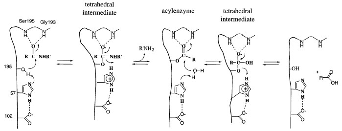 Figure 1. The generally accepted mechanism for serine proteases. (Source: Hedstrom L. 2002)
Types
Macromolecular inhibitors
Macromolecular inhibitors: serpins and small serine protease inhibitors (smapins). The latter group usually acts to irreversibly inhibit enzyme function in a covalent or quasi-covalent manner.
Small-molecule inhibitors
Small-molecule inhibitors: PMSF, DTT, pepstatin, and various plant and animal natural inhibitors (e.g. soybean and lima bean trypsin inhibitors, Bowman–Birk inhibitors from egg white, and aprotinin).
Structural Families
Sources and Biological Roles
Medical and Agricultural Applications
References
Loading ......
