
Inquiry Basket
 Loading ......
Skin biopsy is a procedure in which a small sample of skin is removed for testing. A skin biopsy is used to help diagnose a variety of skin conditions including skin disorders such as psoriasis and eczema bacterial or fungal infections of the skin, and skin cancer. A biopsy can confirm or rule out whether a suspicious mole or other growth is cancerous. This method is performed by excising skin living tissue for histopathological examination, such as H&E staining, immunofluorescence, special staining, immunohistochemistry, etc., which is of great value in diagnosis, classifying, guiding treatment, and prognosis.
Especially for skin cancer, skin cancer including malignant melanoma, basal cell and squamous cell cancer. Basal and squamous cell cancers are more common and treatment effective. Malignant melanoma can be difficult to treat. Early diagnosis and treatment can improve survival in patients with melanoma. A variety of target molecules expressed on skin tissue is used for the clinical diagnosis of skin disease.
At times during a surgical procedure, it is necessary to perform a rapid diagnosis to plan treatment. At critical anatomical sites, a special technique of frozen section control (Mohs surgery) may be used. Mohs surgery is a precise surgical technique used to treat skin cancer. During Mohs surgery, thin layers of cancer-containing skin are progressively removed and examined until only cancer-free tissue remains. This is almost always to allow assessment of the resection margins to check the completeness of excision in cases where the extent of the lesion is not easy to see clinically and when major reconstruction will be required at the time of surgery.
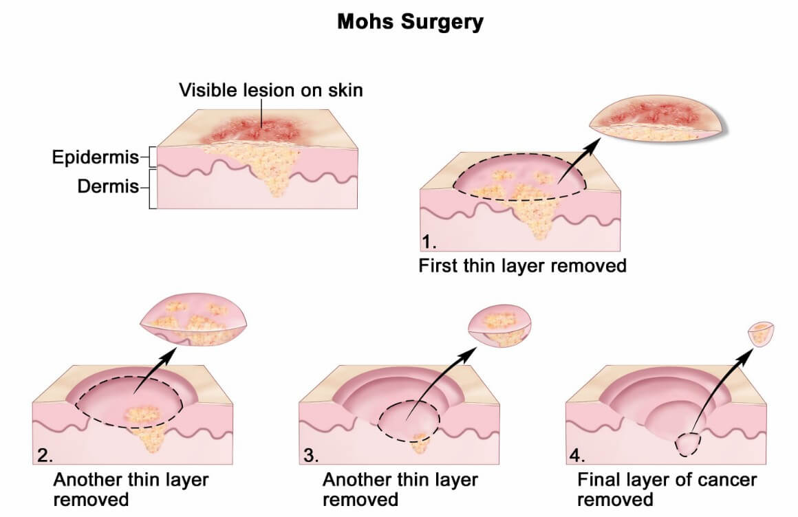 Fig 1. Mohs Surgery
Although the majority of skin biopsies are successfully processed using routine formalin fixation and stained with haematoxylin and eosin (H&E). However Routine H&E staining does not identify all tissue substances in certain inflammatory and neoplastic skin diseases, it may be necessary to stain sections with a variety of different agents or perform different procedures.
Immunohistochemistry is a very powerful tool for identifying cells and substances in tissue sections by using the specificity of antigen-antibody binding. antibodies are raised against particular cellular constituents and these, when applied to a tissue section, will bind specifically to those constituents. The bound antibody is linked by a further step to a colored indicator that can be seen down the microscope (‘staining’).
The major use of immunohistochemistry in skin biopsy is for skin cancer diagnosis, identifying a poorly differentiated malignant tumor as a carcinoma, sarcoma, melanoma, or lymphoma; to identify scanty tumor cells or tumor obscured by inflammation (for instance assessing the depth of a heavily inflamed melanoma), or for identifying a dense lymphoid infiltrate as lymphoma by demonstrating clonality of immunoglobulin light chain expression. The most commonly used antibodies in skin tissue IHC are those raised against cytoskeletal proteins (‘intermediate filaments’) such as cytokeratin, desmin and actin, and markers of melanocytic lineage or differentiation. There are also numerous markers of lymphoid differentiation. Melanocytic differentiation markers such as S100, HMB-45, Melan-A; Lymphoid / Haematological markers such as CD45, CD3, CD4/CD8, CD20/CD79a, CD30, CD68 etc.
Direct immunofluorescence staining is a specific type of immunohistochemistry, usually to detect the presence of immunoglobulins and complement in the skin. Fluorescein-labeled antibody against IgG, IgM, IgA, fibrin, and C3 is applied to frozen sections of fresh tissue and examined by fluorescence microscopy. Another way is detecting circulating skin antibodies by indirect immunofluorescence, in which technique, the patient's serum is incubated with squamous epithelium, and any antibodies that bind are identified by a fluorescein-labeled probe.
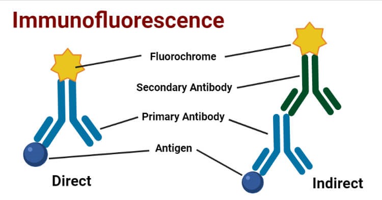 Fig 2. Immunofluorescence
In some cases, identifying specific chromosomal alterations can confirm a diagnosis, which is particularly useful in cutaneous lymphomas. Additionally, this method can further classify the diagnosis, or help differentiate between systemic or primary cutaneous disease. The commonly used technique is fluorescent in situ hybridisation (FISH) where labeled DNA probes attach known areas on specific chromosomes. Equally, immunohistochemistry can be used to stain proteins gained or lost from these mutations.
Electron microscopy (EM) is rarely required for routine diagnosis and management of skin diseases. Scanning EM is useful for diagnosing rare genetic hair shaft abnormalities. It can be combined with immunohistochemistry techniques for working up certain cases, such as for the study of genetic and autoimmune bullous diseases.
Loading ......
