
Inquiry Basket
 Loading ......
Rabies virus belongs to the family Rhabdoviridae and the genus Rabies virus. The virus particle consists of two parts: a shell and a core. The shell is a tightly packed and complete lipoprotein bilayer envelope, with glycoproteins embedded on the outside and membrane proteins, namely matrix proteins, mainly on the inside. The virus has a spiral shaped nucleocapsid inside, which is composed of single stranded RNA and proteins. The three proteins of nucleoprotein, large transcription enzyme protein or RNA polymerase, and phosphoprotein are collectively referred to as ribonucleic acid proteins.
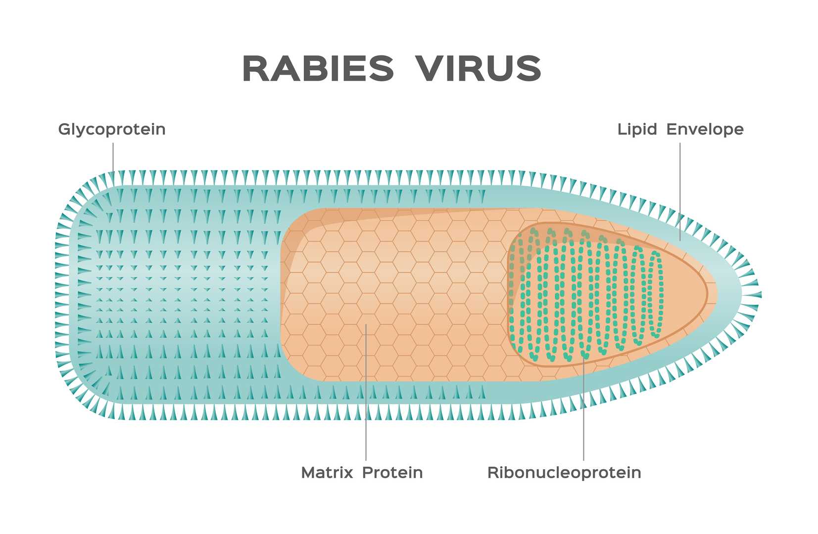 Figure 1. Rabies Virus.
Most animal experiments have shown that viremia does not occur during the incubation period and onset of rabies, and the onset process of rabies can be divided into three stages.
| Pathogenesis | Details |
Propagation period within local tissues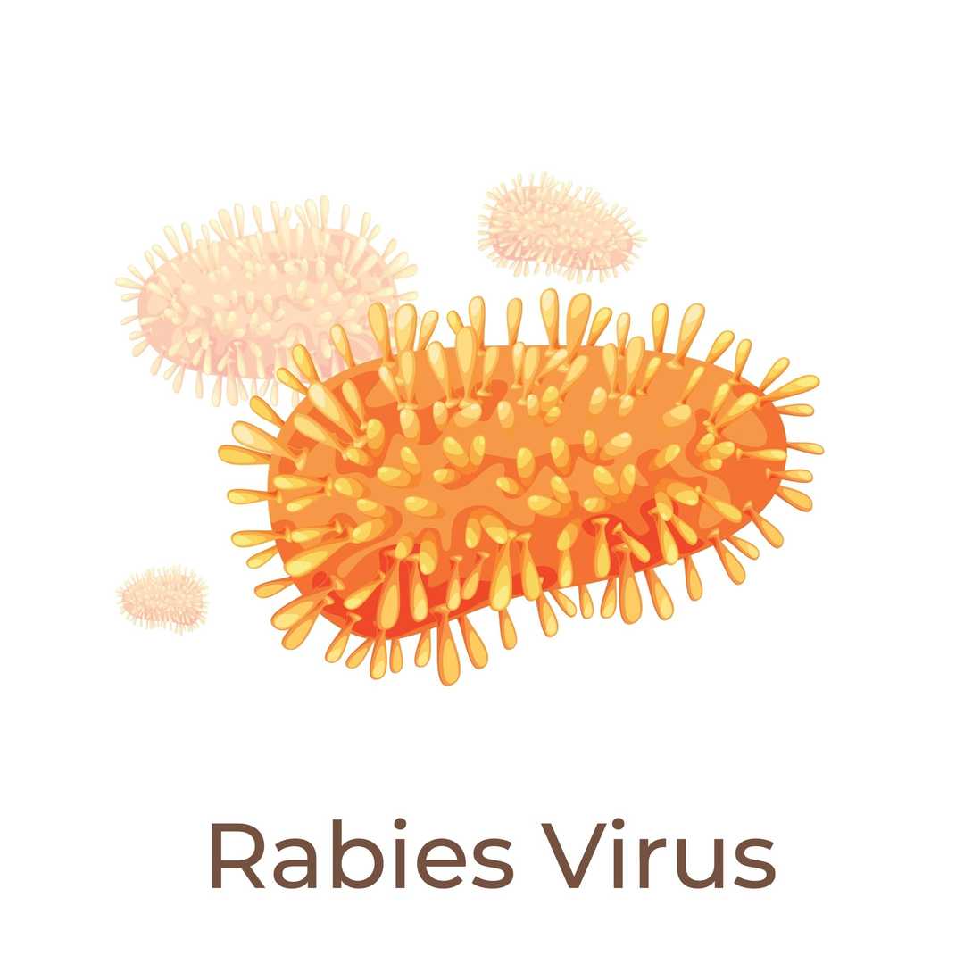 | During the local tissue propagation period, the virus invades the bite site and aggregates and proliferates at the nerve fibers of the striated muscle spindle receptors in the wound. This process involves the binding of the virus G protein to specific receptors on the cell surface, such as nerve cell adhesion molecule (NCAM) or p75 neurotrophic factor receptor (p75NTR), before invading nearby peripheral nerves. |
Invasion of central nervous system phase  | During the invasion of the central nervous system, the virus spreads along the axon cytoplasm of the peripheral nerves towards the heart at a rate of about 3mm per hour. The transport of the virus within the axon relies on its interaction with dynein, which in turn transports it back to the neuronal cell body. |
Spread period to various organs  | During the period of spreading to various organs, the virus undergoes centrifugal diffusion from the central nervous system to the peripheral nerves. This process also utilizes the cellular transport system, such as the forward axonal transport mediated by kinesin, to invade various tissues and organs, especially the salivary nerve nucleu. At this stage, rabies virus glycoprotein (G protein) can specifically bind to presynaptic membrane receptors on the surface of salivary gland cells. |
The pathological changes are mainly acute diffuse encephalomyelitis, especially in the dorsal root ganglia and spinal cord segments corresponding to the bite site, the hippocampus of the brain, and the medulla oblongata, pons, cerebellum, etc. The susceptibility of neurons in these areas is closely related to the distribution of their cell surface specific receptors (such as p75 neurotrophic factor receptor p75NTR, neural cell adhesion molecule NCAM). The formation of endosomes relies on the interaction between viral nucleoproteins (N), phosphoproteins (P), and host cytokines (such as CRABP1, TAX1BP1, etc.), as well as the aggregation of viral RNA. The endosomes are circular or elliptical in shape, with a diameter of approximately 3-10 μ m. It can also be detected in the cone cell layer of the cerebral cortex, spinal nerve cells, posterior horn ganglia, retinal nerve cell layer, sympathetic ganglia, and other locations. The virus efficiently replicates and excretes by specifically recognizing receptors on the surface of salivary gland cells (such as Annexin II), and monocytes infiltrate the glandular tissue.
Humans usually contract rabies after being deeply bitten or scratched by infected animals, and 99% of human rabies cases are transmitted from rabid dogs to humans. Africa and Asia face the heaviest burden of human rabies, accounting for 95% of global rabies deaths. The Americas have largely contained the spread of rabies among dogs, and bats are now the main cause of human rabies deaths in the Americas. Bat rabies has recently become a public health threat in Australia and Western Europe. Death from contact with foxes, raccoons, skunks, jackals, ferrets, and other carnivorous wild hosts is extremely rare in humans. Additionally, it is known that there have been no cases of rabies after being bitten by rodents.
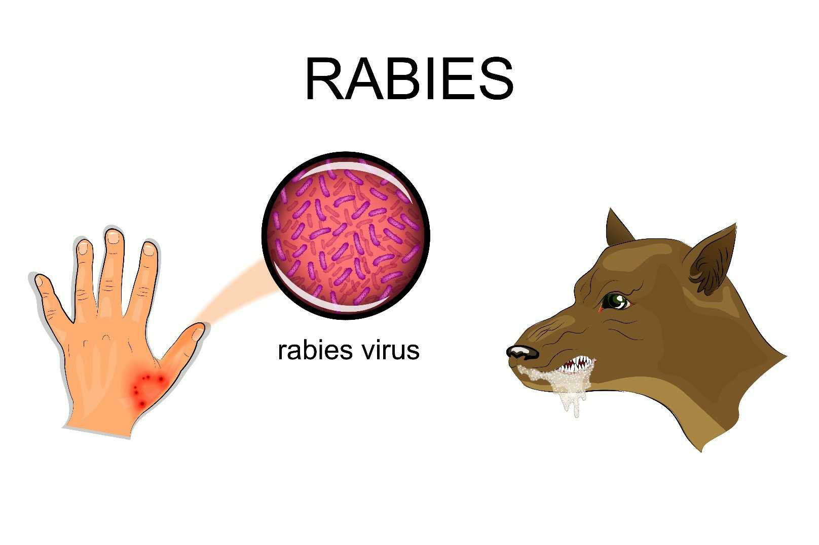 Figure 5. Spread of rabies virus.
The incubation period of rabies is usually 2-3 months, ranging from less than a week to a year, depending on factors such as the location of rabies virus entry and rabies virus load. The initial symptom of rabies is fever, with pain or abnormal or unexplained tremors, stabbing or burning sensations (sensory abnormalities) at the wound site. As the virus spreads in the central nervous system, it develops into lethal progressive brain and spinal cord inflammation.
The symptoms of manic rabies patients are hyperfunction, restlessness, fear of water, and sometimes wind. A few days later, the patient died due to heart and lung failure.
Paralytic rabies accounts for approximately 20% of all human deaths. Compared to manic rabies, its course is less severe and usually longer. Starting from the bite or scratch site, the muscles gradually become paralyzed, and then the patient gradually falls into a coma and eventually dies. Paralytic rabies often has misdiagnosis, resulting in underreporting of rabies.
At present, there is no testing method to diagnose whether a person is infected with rabies before clinical symptoms appear, and if there are no specific signs of rabies such as fear of water or wind, it may be difficult to make a clinical diagnosis. Human rabies can be confirmed in vivo and cadaver through various diagnostic techniques aimed at detecting the whole virus, viral antigens, or amino acids in infected tissues (brain, skin, urine, or saliva).
Wild animals may hide the virus for a long time, so the disease is widely prevalent in wild animals around the world. Foxes, badgers, wolves, mammoths, bats, and other wild carnivores are natural hosts and natural pathogens for the transmission of this disease in nature; In densely populated towns, this disease mainly comes from infected dogs and cats, becoming the main source of transmission for rabies in humans and livestock. Due to the serious harm caused by rabies virus, preventive measures should be taken. Strict management should be implemented for pets such as dogs and cats, and regular vaccination should be administered; If a person is bitten by a rabies dog, the wound should be immediately cleaned and thoroughly washed with 20% soapy water, detergent, amine compounds, or clean water. After cleaning, inject rabies virus immune serum as soon as possible. In addition, scientists are currently studying neurotoxins that can be used to treat diseases caused by viruses such as rabies virus that parasitize the human nervous system.
| Target | Cat. No. | Product Name | Host | Isotype | Application | |
| PRV | CABT-CS517 | Duck Anti-Rabies Virus Polyclonal Antibody | Duck | IgY (∆Fc) | ELISA | Inquiry |
| CABT-NS1217 | Dog Anti-Rabies Virus Glycoprotein Monoclonal Antibody, clone A11 | Dog | IgG2 | WB, ELISA | Inquiry | |
| DMAB-CS23082 | Magic™ Mouse Anti-Rabies Virus Monoclonal antibody, clone 1281 | Mouse | IgG | ELISA (Cap) | Inquiry | |
| DMAB-CS23083 | Magic™ Mouse Anti-Rabies Virus Monoclonal antibody, clone 1282 | Mouse | lgG2a | ELISA (Det) | Inquiry |
| Target | Cat. No. | Product Name | Size | |
| PRV | DAG-WT1123 | Recombinant Pseudorabies Virus gB | 1 mg | Inquiry |
| DAG-WT1124 | Recombinant Pseudorabies Virus gE | 1 mg | Inquiry | |
| DAG-WT1125 | Recombinant Pseudorabies Virus Chimeric Protein (gE&gI) | 1 mg | Inquiry | |
| DAG-WT5927 | Recombinant Rabies virus Glycoprotein Trimer | 100 μg | Inquiry | |
| DAG-WT3649 | Inactivated Rabies virus Culture Fluid | 1 mL | Inquiry | |
| DAGF-021 | Recombinant Rabies virus Glycoprotein (aa 20-458)[His] | 100 μg | Inquiry |
Loading ......
