
Inquiry Basket
 Loading ......
Clinical tumor markers are biomolecules that indicate the presence or progression of cancerous diseases in the body. These markers can include proteins, antigens, genes, and enzymes, typically produced by the tumor itself or the body in response to the presence of cancer. The detection and quantification of these biomarkers provide valuable tools for cancer screening, diagnosis confirmation, prognosis assessment, and treatment monitoring. The clinical application of tumor markers is not limited to detection. Continuous measurement can reveal treatment response and potential recurrence signals long before clinical symptoms or radiological evidence appear.
Enzyme linked immunosorbent assay (ELISA) has become the cornerstone technology for quantitative tumor biomarker detection due to its exceptional sensitivity, specificity, and adaptability to clinical laboratory environments. The quantitative ELISA kit provides values that can be tracked over time to monitor disease progression or treatment efficacy. The quantitative nature of these test kits enables clinicians to establish individual baselines for patients and detect clinically significant deviations that may require intervention. In addition, standardization of cross laboratory ELISA protocols facilitates comparability of results and enables the establishment of a universal reference range for specific tumor markers, thereby enhancing their clinical utility in different patient populations.
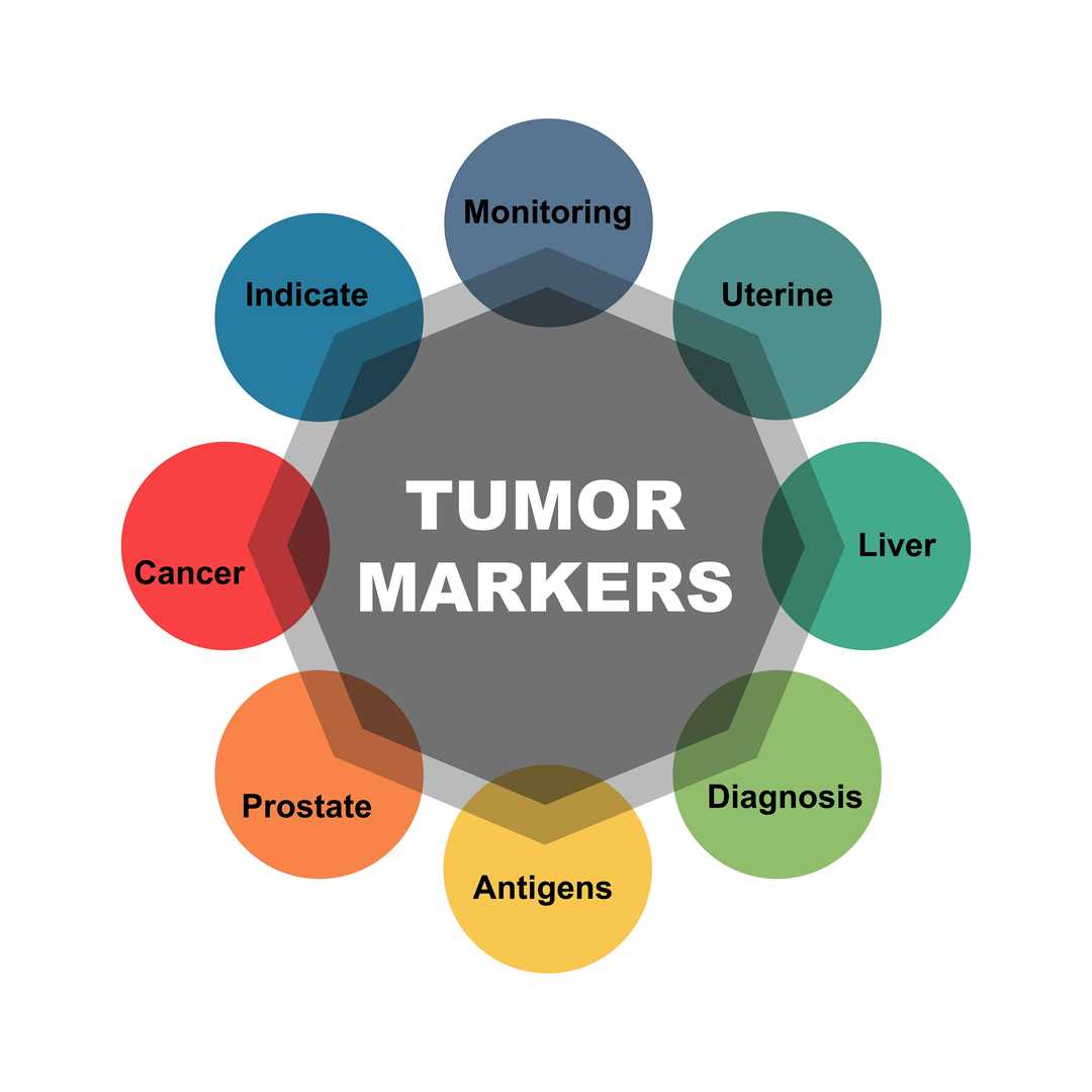
 Figure 1. Cancer of Tumor Markers.
Accurate quantification requires constructing a standard curve by plotting the absorbance value of the calibration standard against its known concentration, and using regression models (such as four parameter logistic regression) for concentration calculation. Key performance indicators include:
Sample Compatibility
Sensitivity Range
Main Uses
The prospects of clinical tumor markers include multiple molecules associated with various types of cancer. The most widely used is carcinoembryonic antigen (CEA), a glycoprotein that increases in colorectal cancer, gastric cancer, pancreatic cancer, lung cancer and breast cancer. Prostate specific antigen (PSA) used for screening and monitoring prostate cancer. Cancer antigen 19-9 (CA19-9) for pancreatic cancer and biliary tract cancer. Cancer antigen 125 (CA125). Alpha fetoprotein (AFP) is used for hepatocellular carcinoma and germ cell tumors. The CA72-4 ELISA kit is a specialized reagent for quantitative detection of tumor markers. The detection range of this kit is usually 0.31-21 ng/mL, covering clinically relevant concentrations for gastrointestinal and ovarian cancer. The design of this kit incorporates specially selected antibodies to minimize cross reactivity with relevant antigens, ensuring reliable measurement of CA72-4 without interference from structurally similar molecules.
Table 1: Selected Clinical Tumor Markers and Their Characteristics
| Tumor Marker | Primary Cancer Associations | Detection Range | Clinical Utility |
| CEA | Colorectal, Lung, Breast, Gastric | Varies by kit | Diagnosis, Monitoring, Recurrence Detection |
| CA19-9 | Pancreatic, Biliary Tract | Varies by kit | Diagnosis, Monitoring |
| CA72-4 | Gastric, Ovarian | 0.31-20 ng/mL | Primary marker for gastric cancer |
| CA15-3 | Small Cell Lung Cancer | Research use | Diagnosis, Treatment Monitoring, Prognosis |
| PSA | Prostate | Varies by kit | Screening, Diagnosis, Monitoring |
Case Study 1: Evaluating the prognostic value of serum p53 levels in colorectal cancer using a quantitative ELISA kit
Sample collection: Collect pre treatment venous blood samples from all participants. Serum was separated by centrifugation and stored at -80 ° C until analysis.
P53 quantification: Measure the serum p53 concentration of all samples using a total p53 (human) ELISA kit. This sandwich ELISA kit is designed to detect wild-type and mutant human p53 protein. The experiment was conducted according to the manufacturer's instructions, using pre coated plates, specific anti-p53 antibodies, and colorimetric detection systems.
Results
Elevated serum p53 in CRC patients: Research has found that the median concentration of serum p53 in the CRC patient group is significantly higher than that in the healthy control group, with a p-value<0.001.
Correlation with disease progression: Serum p53 levels are strongly positively correlated with TNM staging of cancer. The p53 level in late stage (stage III/IV) patients is significantly higher than that in early stage (stage I/II) patients.

Case Study 2: The value of evaluating serum CEA levels in the prognosis and treatment monitoring of colorectal cancer


We collect and process clinical samples (serum/plasma) to eliminate hemolysis/hyperlipidemia and ensure compliance with pre analytical quality standards prior to testing.

We initialize the ELISA kit by preparing calibration samples constructed from standard curves and running low/high concentration quality control materials.

We add the processed sample to a pre coated microplate, incubate to form a sandwich complex, and perform sequential washing to remove non-specific binding.

We measured absorbance using a microplate reader, and calculated tumor marker concentrations using a validated regression model.

We cross validate the results based on the quality control scope, address potential interferences (such as hook effects), and generate clinically actionable reports.
High quality antibodies: We use highly specific monoclonal or polyclonal antibodies with minimal cross reactivity to ensure accurate quantification of your target tumor markers without interference.
Committed to innovation: We continuously invest in research and development, develop new detection methods, and improve existing technologies to ensure that you have access to the most advanced tumor biomarker detection tools.
Customized solutions: In addition to ready-made products, we also provide customized testing and development services - from antibody pair development to full kit production - tailored to your unique research requirements.
Tumor biomarker detection increases multiplexing to capture the complexity of cancer biology, integrates with other omics technologies for comprehensive patient analysis, and automates to enhance reproducibility and facilitate implementation in different medical environments. The quantitative ELISA kit for clinical tumor markers is an indispensable tool in modern oncology, connecting laboratory science with clinical practice through reliable and reproducible measurement of cancer-related molecules. The continuous improvement of these detection methods, which enhances sensitivity, multiplexing capability, and workflow efficiency, is expected to further expand their clinical applications. Clinical decision-making during the treatment process provides critical data.
The selection process should prioritize analyzing performance characteristics, including sensitivity, detection range, accuracy, and specificity, to ensure that they meet clinical requirements. The sample type (serum, plasma, etc.) and required volume should be compatible with your laboratory workflow.
Blood samples should be collected according to standardized protocols, typically using serum separation tubes with appropriate clot formation prior to centrifugation. Hemolysis or lipid samples should be avoided as they may interfere with detection performance. For short-term storage (up to 24 hours), samples can usually be kept at 2-8 ° C, while for long-term storage, it is recommended to freeze at -20 ° C or -80 ° C.
Yes, these platforms use color coded magnetic microspheres coated with different capture antibodies, allowing for mixed detection in one well. While providing comprehensive analysis and sample preservation, multiple detection may have slightly different performance characteristics compared to single analyte ELISA, and should be validated for specific clinical applications.
Explanatory challenges include understanding that elevated levels of most tumor markers do not necessarily indicate malignancy, as benign diseases (inflammation, organ dysfunction, smoking) can also lead to an increase. Similarly, normal levels cannot completely rule out cancer. The dynamic range of analysis must be considered - samples with concentrations exceeding the highest standard need to be diluted and reanalyzed.
Loading ......
