
Inquiry Basket
 Loading ......
Parvovirus is an enveloped, single stranded, DNA virus. Canine animals can be infected through small antigen transfer and natural mutations. Canine parvovirus (CPV) is an acute infectious disease caused by canine parvovirus infection in young dogs. There are two clinical manifestations, hemorrhagic features. The myocarditis type is characterized by sudden death. No matter what type of clinical manifestation, it is characterized by high incidence rate, high mortality and strong infectivity.
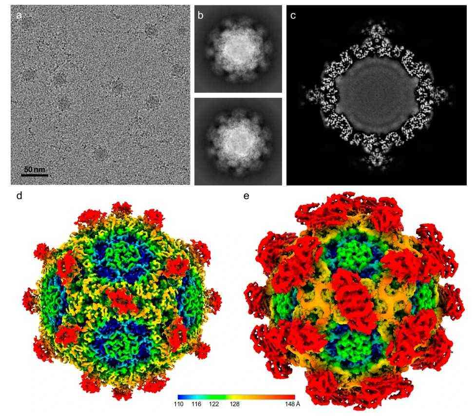 Figure 1. Infectious parvovirus B19 circulates in the blood.( Lee, Hyunwook, et al.2024)
Parvovirus belongs to the genus of viruses, with virus particles having no capsule. CPV has strong resistance to the outside world and is resistant to alcohol, ether, and chloroform. It also has a certain tolerance to temperature. In addition, ultraviolet radiation can also inactivate it. If toxic feces and intestinal tubes are refrigerated in a low-temperature environment, their infectivity can be maintained for a long time. The prevalence of canine parvovirus has no obvious seasonal pattern and can occur throughout the year, but it is more common in winter and spring. Dogs carrying viruses are the main source of infection, and the vomit, feces, and urine of sick dogs all contain a large amount of viruses. There are two ways of transmission: direct and indirect. The former is when healthy dogs without immunity come into direct contact with sick dogs and are infected, while the latter is when healthy dogs without immunity ingest contaminated water, feed, etc. and become infected. This disease mainly affects puppies under half a year old, especially those under three months old. Once infected, more than 50% of the cases are a mixture of gastroenteritis and myocarditis, with a mortality rate of over 80%, and sometimes the infection rate can reach up to 100%.
Analysis of gene sequences of different isolates revealed that CPV is prone to generate new mutant strains through antigen drift, with three variants: CPV-2a, CPV-2b, and CPV-2c. Using MEGA5.0 for comparative analysis of VP2 protein sequences of different CPV strains, it was found that compared with CPV-2, the amino acids of VP2 protein in CPV-2a underwent mutations in Met87Leu, Ile101Thr, Ala300Gly, Asp305Tyr, and Val555lle; Compared with CPV-2a, CPV-2b has an additional Asn426Asp amino acid mutation in the VP2 protein.
Canine parvovirus belongs to the Parvoviridae family, Parvovirus genus, and is an enveloped carnivore virus. The nucleotide homology between CPV and small viruses such as feline panleukopenia virus (FPLV) and mink enteritis virus (MEV) exceeds 98%. CPV is the smallest linear negative stranded single stranded DNA virus, with a genome length of 5323 bp. It consists of a 5000 nucleotide DNA molecule and contains two coding boxes, encoding non structural proteins NS1 and NS2, as well as structural proteins VP1 and VP2. CPV exhibits the typical morphology and structure of the Parvovirus genus: the virus particle is an icosahedron with equiaxed symmetry and no envelope. Under electron microscopy, it appears circular or hexagonal, with a diameter of approximately 26 nm. The nucleocapsid is composed of 32 3-4 nm-long capsomeres, and the entire virion is mainly formed by 60 alternating motifs of VP1 and VP2. As the major capsid protein, VP2 is critical for viral attachment to host cells and immunogenicity—Recombinant CPV VP2 Protein (mimicking the native VP2 structure) has become a key tool for studying viral tropism and developing diagnostic assays. Additionally, Anti-CPV monoclonal antibody, which specifically recognizes VP2 epitopes, enables precise detection of viral particles in clinical samples and facilitates research on CPV's pathogenic mechanism, such as its interaction with host cell receptors.
Dogs develop symptoms within 3-14 days after exposure to parvovirus, with an average onset time of 5-7 days. Clinical symptoms include decreased appetite, depression, fever, vomiting, diarrhea, and a foul odor of canine parvovirus in feces. The incidence rate of adult dogs (dogs more than 12 months old) is very low. If the dogs are not of high purity or closely mated adult dogs, there will be no obvious symptoms. It only shows that the feces have a certain odor, and appetite decreases. If there is no aggravation, there is no need for treatment. Dogs infected with canine parvovirus can be clinically classified into enteritis type and myocarditis type.
| Classification | Details |
| Enteric | The incubation period of natural infection is 7 to 14 days. At the beginning of the disease, the symptoms include fever, body temperature can reach over 40 ℃, mental depression, refusal to eat, and vomiting. The vomitus in the initial stage was undigested food and white foam (stomach acid), followed by mucoid and yellowish green liquid. Diarrhea begins one day after the onset of the disease. At the beginning of the illness, the feces are thin or semi shaped, with a certain fishy odor. As the condition progresses, the stool becomes a dark green or yellow jelly like substance. |
| Myocarditis Type | Commonly seen in puppies around 40 days old, sick dogs have no obvious clinical symptoms, some suddenly have difficulty breathing, heart failure, and die in a short period of time; Some sick dogs die after mild diarrhea. Canine parvovirus myocarditis is caused by the attack of canine parvovirus on the cardiac muscle cells of dogs, which can damage the cardiac muscle cells of dogs in a short period of time. |
CPV spreads through the fecal oral route, attacking host intestinal epithelial cells and myocardial cells. The virus first replicates in the lymphoid tissue of the oropharynx, mesenteric lymph nodes, and thymus, and then spreads throughout the body under the influence of blood circulation. Within 3-5 days, it can cause viremia and then spread to the villous crypts of the small intestine, forming eosinophilic nuclear inclusions. When CPV infects myocardial cells, myocardial capillaries undergo dilation and congestion, while lymphoid and bone marrow proliferating cells are severely damaged, leading to a decrease in lymphocytes. In the small intestine of healthy dogs, intestinal epithelial cells that migrate from the crypt end differentiate and mature at the top of the villi, and then acquire the ability to digest and absorb nutrients, thereby helping the body absorb nutrients. But after CPV invades the intestinal crypts, the renewal of normal intestinal epithelial cells is disrupted, and the formed villi become dull, shrink, and gradually lose their absorption capacity. Severe enteritis can lead to self poisoning due to the absorption of endogenous toxins, as well as an increased risk of viral translocation and Escherichia coli sepsis, resulting in septic shock and ultimately death. Research has found that in 2-3 week old serum negative puppies, CPV can replicate in myocardial cells, causing acute myocarditis.
| Immunoassay Options | Details |
| Colloidal gold immunochromatographic assay (GICA) | It is a solid-phase labeling immunoassay technology that combines gold labeling, immunoassay, and liquid chromatography techniques. It can quickly and easily diagnose CPV and is therefore commonly used in primary clinical testing. |
| Polymerase Chain Reaction (PCR) | PCR method is the main method for detecting CPV due to its high sensitivity, strong specificity, and easy operation. Currently, multiple PCR methods have been developed. |
| Enzyme linked immunosorbent assay (ELISA) | It is the most commonly used testing method by clinical doctors and practitioners for detecting CPV from the feces and blood of infected dogs. ELISA is used for detecting and quantifying antigens and target specific antibodies, and can detect a large number of samples at once. Various reagent kits have been developed domestically and internationally for clinical use, but false negative and false positive results are prone to occur. |
References
Loading ......
