Extracellular vesicles (EVs) are small, membrane-bound particles released by cells into the extracellular environment. These vesicles contain a variety of bioactive molecules, including proteins, nucleic acids, and lipids. These molecules can be transferred to recipient cells and modulate various physiological and pathological processes. EVs have garnered significant attention in biology due to their role in intercellular communication and their potential as biomarkers for various diseases.
Both small and large EVs can be released from normal healthy cells under physiological conditions to ensure intercellular communication, thereby regulating a range of biological processes from cell maintenance to immune responses. Due to the difference in diameter and mode of occurrence, there are three main types of EVs: exosomes, microvesicles, and apoptotic vesicles.
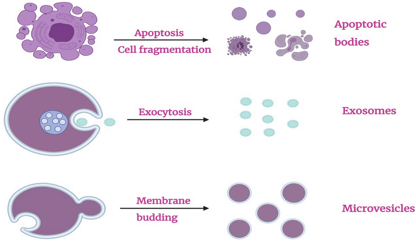 Fig. 1 Types of extracellular vesicles (EVs). (Alghamdi M, et al., 2022)
Exosomes are derived from the endocytic pathway of cells. They are typically small in size, ranging from 30 to 150 nanometers in diameter, and are known for their unique lipid bilayer membrane composition. Exosomes can be found in various biological fluids, including blood, urine, and saliva, making them an attractive source for biomarker discovery. These vesicles are released by both healthy and diseased cells, and their cargo can reflect the physiological or pathological state of the parent cell.
Microvesicles, also known as microparticles, are larger than exosomes and range in size from 100 to 1,000 nanometers. Unlike exosomes, microvesicles are released by a process known as budding or shedding from the plasma membrane of cells. They have a more heterogeneous composition, containing not only proteins and nucleic acids but also cellular organelles and membrane receptors. Microvesicles have been implicated in various physiological processes, such as coagulation, immune responses, and tissue repair.
Apoptotic bodies are EVs released during programmed cell death, specifically apoptosis. When a cell undergoes apoptosis, it undergoes structural changes, including the formation of apoptotic bodies. These EVs contain numerous cellular components, including fragmented DNA and cytoplasmic organelles. Apoptotic bodies play a crucial role in the efficient removal of dying cells by phagocytic cells.
The isolation of EVs from complex biological samples presents a significant challenge due to their small size and heterogeneity. Various methods have been developed to overcome these challenges and facilitate downstream EV analysis. One widely used technique is differential ultracentrifugation, which involves sequential centrifugation steps to isolate EVs based on their size and density. Other methods include size-exclusion chromatography, ultrafiltration, and immune affinity capture by specific antibodies against EV surface markers. Once isolated, various techniques can be used for the analysis of EVs, including Western blotting, ELISA, and mass spectrometry. These techniques enable the detection and quantification of various molecules in EVs, including proteins, lipids, and nucleic acids.
Biomarkers play a pivotal role in disease detection, prognosis, and therapeutic response monitoring. Traditional biomarkers, such as proteins or nucleic acids, may be limited in their ability to accurately represent the complexity of diseases. This is where extracellular vesicles come into play. These tiny structures carry specific molecular signatures reflecting the physiological or pathological state of the parent cells. Therefore, the analysis of extracellular vesicles holds substantial promise for biomarker discovery and precision medicine.
EVs have been shown to be involved in major physiopathological processes, including cellular homeostasis, infection propagation, cancer development, and cardiovascular disease. More and more studies have found that EVs have many advantages over traditional synthetic carriers, opening up new fields for modern drug delivery.
As drug delivery vehicles, EVs have the following numerous advantages:
Based on these unique advantages of extracellular vesicles, EVs secreted by 293 cells, mesenchymal stem cells (MSCs) and other cells have been developed by researchers into small molecule drugs and nucleic acid drug carriers for therapeutic research in diseases of the respiratory system, tumors, the central nervous system, and cardiovascular diseases.
Reference