
Inquiry Basket
 Loading ......
The autoimmune ELISA kit for the nervous system is an important tool in modern neuroscience research, which can accurately detect and quantify autoantibodies targeting neural antigens. These kits utilize enzyme-linked immunosorbent assay (ELISA) technology to provide reliable quantitative data on autoimmune responses targeting key neural structures. Accurately measuring neural autoantibodies helps to study the mechanisms of neuroinflammation, disease progression, and the efficacy of treating multiple sclerosis, neuromyelitis spectrum disorder (NMOSD), autoimmune encephalitis, and other autoimmune mediated neurological diseases. These ELISA kits target specific autoantibodies, including anti myelin oligodendrocyte glycoprotein (MOG), anti myelin basic protein (MBP), anti-aquaporin-4 (AQP4) and other nervous system autoantibodies, which have significant diagnostic and research value in basic science research and translational studies.
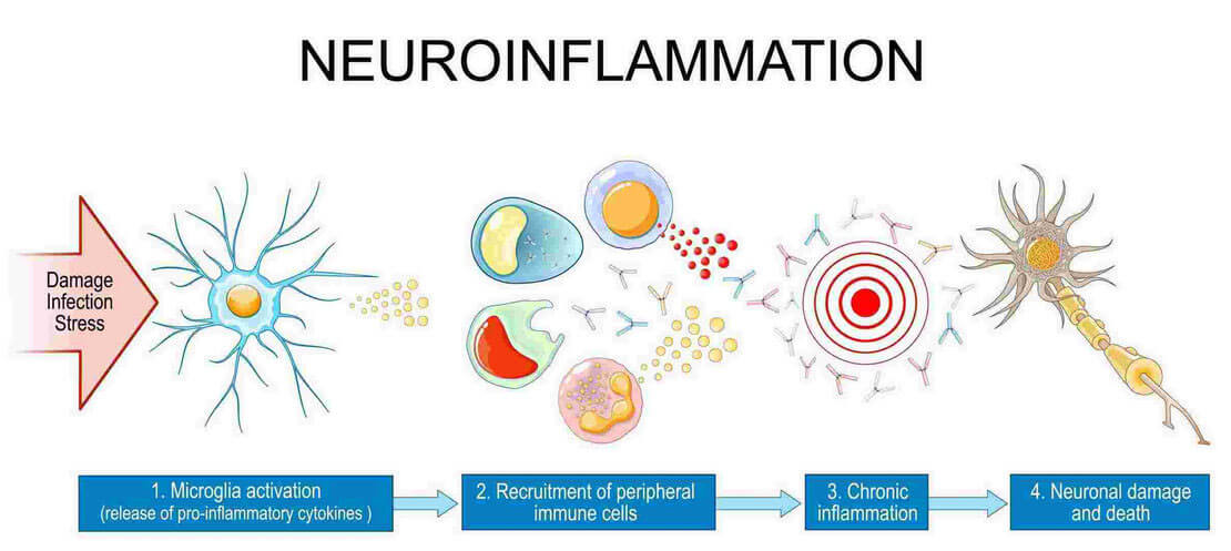 Figure 1. Neuroinflammation and Autoimmune.
The autoimmune response of the nervous system involves complex immune-mediated processes that target specific components of the nervous system, leading to functional impairment and potential systemic consequences. The pathogenesis of autoimmune neurological diseases usually begins with the breakdown of immune self tolerance, followed by the production of autoantibodies against neuroantigens. These autoantibodies may directly interfere with neurological function through receptor blockade or stimulation, or serve as markers of sustained autoimmune activity. The relationship between the autoimmune response of the nervous system and systemic inflammation is an important research field. Autoimmune nervous system diseases often occur simultaneously with other autoimmune diseases, resulting in complex clinical manifestations that require comprehensive diagnostic methods. For example, studies have shown that anti nuclear antibodies (ANA) traditionally associated with rheumatism can also bind to neuronal targets, which may lead to neurological symptoms in diseases such as neuropsychiatric lupus erythematosus.
 Figure 2. Neuroinflammation and Autoimmune Research.( Bardehle, Sophia, et al. 2017)
Neurological autoantibody markers are highly specific immunoglobulins that target antigens within the nervous system, serving as indicators of autoimmune activity and potential neurological dysfunction. These biomarkers vary in pathological significance, diagnostic specificity, and clinical application:
Anti-Myelin Oligodendrocyte Glycoprotein
These antibodies target MOG, a protein expressed on the outermost surface of the myelin sheath in the central nervous system. MOG antibodies are mainly associated with MOG antibody related diseases (MOGAD).
Anti-Myelin Basic Protein (MBP)
These autoantibodies target MBP, which is the main component of myelin sheath. MBP contains approximately 30% myelin protein and plays a crucial role in maintaining myelin structure through interactions with lipid bilayers.
These antibodies target aquaporin 4, which is highly expressed at the end of astrocytes in the blood-brain barrier. The AQP4 antibody has high specificity for neuromyelitis spectrum disorder (NMOSD) and is detected in approximately 70-80% of patients.
Anti-N-Methyl-D-Aspartate Receptor (NMDAR)
NMDAR antibodies are associated with anti NMDAR encephalitis, characterized by neuropsychiatric syndromes that may include psychiatric symptoms, seizures, memory deficits, autonomic instability, and decreased levels of consciousness.
Neurological autoimmune ELISA kits serve diverse applications across basic research, preclinical studies, and therapeutic development:
Biomarker discovery and validation
The quantitative ability of ELISA system is helpful for the identification and validation of biomarkers in neuroautoimmunity. By measuring the levels of autoantibodies in a patient population and correlating them with clinical features, imaging results, and other laboratory parameters, researchers can identify potential biomarkers for diagnosis, prognosis, or treatment response prediction.
Treatment monitoring and clinical trials
These ELISA kits provide objective endpoints for evaluating new therapies for autoimmune neurological diseases. Researchers use a series of autoantibody measurements to evaluate treatment efficacy, identify target populations, and monitor the potential immunogenicity of biological therapies.
Disease modeling and characterization
In experimental models such as autoimmune encephalomyelitis (EAE), ELISA kits enable researchers to quantify humoral responses to specific neuroantigens such as MOG and MBP. This enables the characterization of autoimmune responses in different disease models and the evaluation of their correlation with clinical course and neuropathological outcomes.
When combined with complementary research methods, the nervous system autoimmune ELISA kit is the most powerful:
| Integration | Details |
 | Correlation with clinical phenotype Combining serological data with detailed clinical assessments enables researchers to establish meaningful correlations between autoantibody profiles and disease characteristics. |
 | Integration with neuroimaging Combining ELISA results with advanced neuroimaging techniques can provide a deeper understanding of the structure and functional consequences of the nervous system's autoimmune response. |
 | Supplementary molecular techniques ELISA data can be enhanced through integration with other laboratory methods, including cell-based detection, immunohistochemistry, and molecular biology techniques, to comprehensively understand autoimmune mechanisms. |
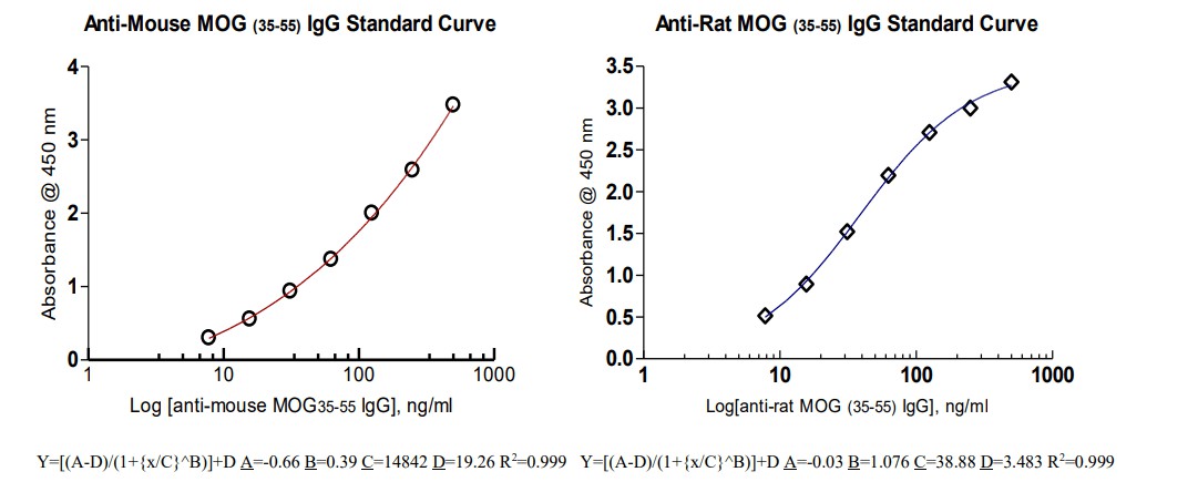
Case Study 1: Anti-MOG (35-55) IgG ELISA Kit in Experimental Autoimmune Encephalomyelitis Research
Background: Myelin oligodendrocyte glycoprotein (MOG) is an important target antigen for autoimmune demyelinating diseases, and MOG antibodies are associated with human diseases and experimental models. The performance of the anti-MOG (35-55) IgG ELISA kit in detecting and quantifying anti MOG antibodies in experimental autoimmune encephalomyelitis (EAE), a mature model for multiple sclerosis research, was evaluated.
Experimental methods and results: The quantitative properties of ELISA indicate that higher concentrations of anti MOG antibodies are significantly correlated with clinical disease scores and neuropathological findings, including demyelination and immune cell infiltration. This measurement demonstrates excellent accuracy with appropriate intra - and inter batch coefficients of variation, confirming good reproducibility across measurements.
Case Study 2: Rat MBP (Myelin Basic Protein) ELISA Kit in Demyelination Research
Background: Myelin basic protein (MBP) is the main protein component of myelin sheath, accounting for approximately 30% of myelin protein in the central nervous system. MBP plays a crucial role in the structure and stability of myelin sheaths, and anti MBP antibodies have been studied under various demyelinating conditions. We evaluated the performance of the rat MBP ELISA kit in detecting and quantifying MBP in research applications, with a particular focus on its practicality in demyelination models.
Experimental methods and results: The evaluation used a rat MBP ELISA kit, which included samples from demyelination and myelin regeneration experimental models. This assay demonstrates appropriate sensitivity for detecting MBP in various sample types, including tissue homogenates and possibly other biological fluids. In pathological research, MBP concentration is correlated with the degree of demyelination and myelin regeneration, supporting the biological relevance of quantitative MBP measurement.


We collect and process clinical samples (serum/plasma) to eliminate hemolysis/hyperlipidemia and ensure compliance with pre analytical quality standards prior to testing.

We initialize the ELISA kit by preparing calibration samples constructed from standard curves and running low/high concentration quality control materials.

We add the processed sample to a pre coated microplate, incubate to form a sandwich complex, and perform sequential washing to remove non-specific binding.

We measured absorbance using a microplate reader, and calculated tumor marker concentrations using a validated regression model.

We cross validate the results based on the quality control scope, address potential interferences (such as hook effects), and generate clinically actionable reports.
The autoimmune ELISA kit for the nervous system is an indispensable tool for advancing research on neuroinflammation and autoimmune diseases. These complex immunoassays provide researchers with the critical ability to detect and quantify specific neural autoantibodies and antigens with extremely high precision and reliability, fundamentally enhancing our understanding of the autoimmune pathogenesis that affects the nervous system. The implementation of these standardized ELISA systems has significant advantages throughout the entire research field. These test kits can comprehensively study disease mechanisms, facilitate autoantibody analysis for disease modeling, and support the evaluation of new therapies through quantitative monitoring of autoimmune responses and myelin dynamics.
MOG antibodies are associated with a unique clinical entity called MOG antibody related disease (MOGAD), which includes phenotypes such as optic neuritis, transverse myelitis, acute disseminated encephalomyelitis (ADEM), and single or multiple focal defects in the brain. Unlike multiple sclerosis, MOGAD typically exhibits different treatment responses and prognoses, so accurate antibody testing is crucial for proper management.
Compared with other methods such as cell-based detection (CBA) or immunohistochemistry, ELISA systems provide good performance, quantitative results, and relatively simple implementation. Although CBA is often considered the gold standard for certain autoantibodies such as AQP4 and MOG, as they can detect conformationally sensitive antibodies, ELISA platforms provide a good balance of performance, practicality, and throughput for many research applications.
Most test kits have been validated and can be used with serum or plasma collected using standard techniques. Depending on the specific kit, some tests can also accommodate other sample types, such as tissue homogenates or possibly cerebrospinal fluid (CSF). Proper sample processing is crucial for maintaining the integrity of autoantibodies and obtaining reliable results.
Yes, quantitative autoantibody measurement can provide valuable information about treatment response in research environments. For example, a decrease in MOG antibody levels has been observed in some patients receiving immunotherapy, although the clinical significance of continuous monitoring varies depending on autoantibodies. Similar patterns also exist in other autoimmune neurological diseases, although biological and clinical relevance remain crucial.
Reference
Loading ......
