
Inquiry Basket
 Loading ......
Myoglobin (MB) belongs to the globin superfamily and has been of great interest since its discovery. The essential functions of the globin superfamily include sensing of oxygen (O2), detoxification of reactive oxygen species (ROS), reactive nitrogen species (RNS), and other reactive species, and functional coupling to O2-dependent enzymes. In addition, several other gas molecules similar in size to O2, such as nitric oxide (NO) and carbon monoxide (CO), are able to enter the body through similar protein channels and interact with the heme moiety.
Table 1. Binding affinities and reduction rates for globin proteins
| Globin | NO binding dissociation constant | O2 binding affinity | Nitrite reduction rate, M-1 Y-1 |
| Hemoglobin (tetramer) | 2.5×10⁻⁴ M [Fe(III)] | 26 Torr | 0.12-6 |
| α-Globin | 1.4×10⁻⁴ M [Fe(III)] | 12×1014 M-1 | 9.2 |
| β-Globin | 2.3×10⁻⁴ M [Fe(III)] | 14×1014 M-1 | 10.3 |
| Myoglobin | 0.7×10⁻¹¹ M | 2-3 Torr | 2.9-5.6 |
| Cytoglobin | (S–S) 0.2 Torr (S–H) 2.0 Torr | 0.14 | |
| Neuroglobin | (S–S) 1.0 Torr (S–H) 10 Torr | Mouse: 5.1 Human: (S–S) 0.12 (S–H) 0.062 |
(Source: Keller TCS 4th, et al. 2022)
Initially MB was thought to be expressed only in rhabdomyosin, red skeletal muscle, and cardiac myocytes. Changes in myoglobin expression levels throughout embryonic life up to adulthood show this tissue-restricted expression pattern. Myoglobin concentrations were higher in oxidized muscle fibers than in fast muscle fibers, and MB-encoded DNA sequence transcripts were 333-fold and 25-fold higher in cardiac muscle than in normal colon and breast tissue, respectively. MB is also expressed in metabolically active non-muscle sites, e.g., hypoxia upregulates MB transcript expression more in liver than in muscle. Some researchers found that MB was present in different tissues of common carp and zebrafish, such as liver, brain, kidney, gills, intestine and eyes, with different tissue-dependent MB expression patterns.
MB is predominantly found in the myoplasm, and the three-dimensional structure viewed under the microscope is that of a flattened, spindle-shaped, spherical protein composed of heme cofactors and bead proteins. The amino acids in the side chain of the MB hydrophilic group are almost distributed on the outer surface, the hydrophobic group wrapped around the interior protects heme from oxidation by the external environment and allows the central heme to bind oxygen, and the outer bead protein chain imparts water solubility to the heme moiety so that the protein maintains its function. Heme consists of four pyrrole substituents and an iron atom in the center, and the resonance properties of the conjugated double bond in this group determine the ability of MB to absorb visible light and thus perform its role as a pigment. MB itself is purple in color, and when it comes into contact with oxygen in the air, it will rapidly covalently combine with oxygen to form bright red oxygenated MB, which has a bright red appearance. With increasing oxidation time, Fe2+ in the heme cofactor is oxidized to Fe3+ and MB or MbO2 becomes methemoglobin (Met-MB).
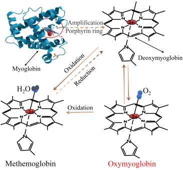 Figure 1. Structural changes of myoglobin
(Source: Han J, et al. 2024)
MB expression profiles are closely related to their functions. MB has been found to remove NO, ROS and induce mitochondrial fusion in addition to its functions of oxygen storage, transport and promotion of intramuscular diffusion.
MB is found primarily in red muscle and provides the oxygen needed to produce the energy required for contraction. MB has the ability to maintain a high affinity for oxygen as well as a high storage capacity for oxygen, allowing oxygen to be taken from the blood and supplied to cytochrome c oxidase. These features can also explain tissue distribution patterns of MB, such as why myoglobin is absent from the fast-beating heart and insect wing muscles, but present in the slow-beating heart, diaphragm, and running muscles that require constant repetitive activity. Another major function of the MB is to participate in intracellular oxygen transport and to facilitate oxygen diffusion, a function that was first thought to be impossible because of the lack of knowledge at the time of the mechanisms involved in circulating or transporting oxygen in such a small space within the muscle fiber. The researchers then proposed that MB facilitates the cellular transport of oxygen throughout the process from the capillary wall to the mitochondria. The oxygen pressure gradient from the capillary to the muscle cell is discontinuous. In addition, the oxygen pressure gradient within the sarcoplasm is shallow, whereas the oxygen pressure gradient at the interface between the capillary wall and the sarcoplasm is large. This steep gradient allows for a continuous inflow of oxygen to the erythrocytes even when delivery is discontinuous.
Although the diffusion rate of free oxygen is 20-fold higher than that of oxygen bound to MB, deoxygenated MB has a high affinity for oxygen, rapidly traps oxygen and diffuses it to the mitochondria, where it is expelled and oxygen homeostasis is maintained. In addition, the concentration of MB-bound oxygen in muscle cells is approximately 30 times that of free oxygen. Based on these phenomena, it has been proposed that three conditions must be met in order for the oxygen flux produced by facilitated diffusion to exceed the flux of free dissolved oxygen. They are, respectively, that the concentration of MB must be quite high and partially desaturated with oxygen somewhere in the system, and that it must be free to undergo translational diffusion within the sarcoplasm. In this way, when entering the sarcoplasm from the sarcolemma, the rate of decrease in the oxygen gradient decreases in parallel with an increase in MB-facilitated oxygen diffusion, which gradually produces free or unsaturated MB. These MBs must be combined with new oxygen to repeat the cycle, thus maintaining oxygen flux.
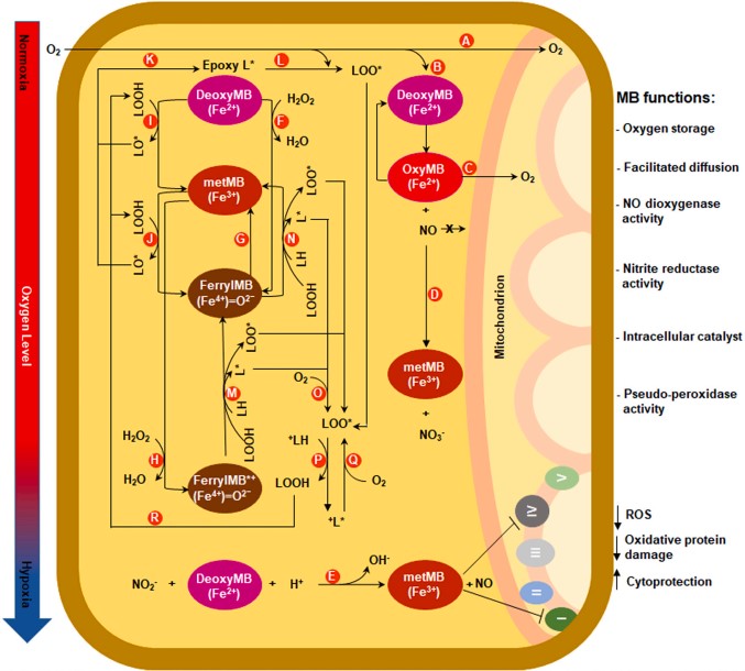 Figure 2. Schematic illustration of different functions of MB
(Source: Elkholi IE, et al. 2022)
MB is upregulated in organisms that are exposed to hypoxic conditions or require increased oxygen levels/supply. The molecular mechanisms behind this regulation are unknown, and hypoxia-inducible factor (HIF-1α) has been found to be a possible regulator of MB in some mouse experiments. HIF-1α is mainly regulated by cellular oxygen levels, and muscle exercise induces HIF-1α to compensate for increased oxygen demand even in the absence of hypoxia. However, the standard promoter of the MB gene lacks a hypoxia-responsive element (HRE) and the 10Kb region upstream of the gene lacks a HIF-1α-binding motif, suggesting that the standard muscle MB transcript is neither directly regulated by hypoxia nor dependent on HIF-1α. The researchers demonstrated that hypoxia requires another stimulus to release calcium from the sarcoplasmic reticulum, which initiates the calcium/calmodulin/calcineurin pathway. In the latter pathway, calcineurin dephosphorylates nuclear factor of activated T-cells (NFAT), leading to its translocation in the nucleus and thus regulating MB transcription.
Myoglobin acts as an NO scavenger under normoxic conditions and as a nitrite reductase under hypoxic conditions, and this dual role allows MB to couple ambient oxygen tension and smooth muscle tone via NO kinetics. NO scavenging mediated by MB may limit the ability of NO to activate secondary messengers that signal smooth muscle relaxation. In addition, MB may control circulatory hemodynamics by preventing smooth muscle relaxation, particularly in the surrounding large conduit arteries.
When attempts were made to disable MB, one investigator found that no effects on cardiac or exercise function were observed in animals with overall actin deficiency, yet another trial observed functional changes in cardiac output, fractional shortening, and respiratory exchange ratios in a mouse MB knockout model. Thus, functional differences may not be strictly related to the oxygen buffering role of myoglobin. In vascular smooth muscle, myoglobin and nitrite are thought to mediate the hypoxic vasodilatory response. Genetic deletion of myoglobin reduces hypoxic vasodilation by half.
Vascular myoglobin promotes circulatory homeostasis by activating NO signaling via nitrite reduction and inactivating NO via deoxygenation. These responses are dependent on myoglobin oxygenation, which positions myoglobin as a specific regulator of tissue perfusion by coupling vasodilatory NO signals to local hypoxic signals.
MB expression in cancer was initially associated only with rhabdomyosarcoma-like differentiation of cancerous tissue and was considered a marker of rhabdomyosarcoma cell differentiation. MB expressed in cancers of non-muscle origin is derived from surrounding muscle tissue, for example, the investigators found that MB mRNA and protein levels were highest in breast cancer and lowest in osteosarcoma, but were not detected in normal breast cell lines or tissue samples. Interestingly the presence of MB has also been reported in the remaining types of cancers without muscle tissue differentiation. However, the mechanisms by which MB is widely expressed or plays a functional role in different cancers remain unclear.
MB has different potential roles in the metabolic processes of many types of cancerous tissues. In liver tissues, overexpression of MB in vitro and in vivo increased ATP levels and attenuated hepatic ischemia-reperfusion injury. MB-transfected A549 human lung cancer cells showed high oxygen content, HIF-1 downregulation, promotion of differentiation, inhibition of angiogenesis, tumor growth and metastasis. In vitro overexpression of MB in MDA-MB-231, MDA-MB-468, and MCF7 breast cancer cells induces mitochondrial hyperfusion, reduces cell proliferation, and further reduces tumor volume in MB-expressing xenografts in vivo.
Table 2. Summary of the reported functions of MB in cancer
| Tumor lineage (cell line) | Manipulation | Molecular consequences | Functional consequences |
| Liver -Hep3B | Overexpression (transient) | Increased ATP level | Not applicable |
| Lung -A549 | Overexpression (stable) | Better oxygenation levels under hypoxia | Decreased primary tumor and metastatic burden |
| Breast -MDA-MB-231 -MDA-MB-468 -MCF7 | Overexpression (stable) | Mitochondrial hyperfusion leading to cell cycle arrest | Decreased proliferation in vitro and MDA-MB-231 tumor burden in vivo |
| -MDA-MB-468 | Knockdown (stable) | Improved oxygenation and higher mitochondrial activity | Decreased proliferation and migration |
(Source: Elkholi IE, et al. 2022)
References
| Target | Cat. No. | Product Name | Size | Species | Application | Detection Sample | |
| MB | DEIA1870 | Human Myo(Myoglobin) ELISA Kit | 96T | Human | Quantitative | Serum, plasma, tissue homogenates and other biological fluids | Inquiry |
| DEIA6155 | Mouse MYO(Myoglobin) ELISA Kit | 96T | Mouse | Quantitative | Serum, plasma, tissue homogenates and other biological fluids | Inquiry | |
| DEIA6150 | Rat MYO(Myoglobin) ELISA Kit | 96T | Rat | Quantitative | Serum, plasma, tissue homogenates and other biological fluids | Inquiry | |
| DEIA6151 | Pig Myoglobin ELISA Kit | 96T | Pig | Quantitative | Serum, plasma, urine | Inquiry | |
| DEIA6152 | Rabbit MYO(Myoglobin) ELISA Kit | 96T | Rabbit | Quantitative | Serum, plasma, tissue homogenates and other biological fluids | Inquiry | |
| DEIA8087 | Human Myoglobin ELISA Kit | 96T | Human | Quantitative | Biological samples | Inquiry | |
| DEIACL22 | CDSimple™ Myoglobin Chemiluminescent ELISA Kit | 96T, 192T | Quantitative | Serum | Inquiry |
| Target | Cat. No. | Product Name | Expression System | Tag/Conjugate | Application | |
| MB | DAGA-861 | Bovine myoglobin (>98%) | Bovine | Unconjugated | ELISA, WB | Inquiry |
| DAGA-862 | Canine myoglobin (>98%) | Dog | Unconjugated | ELISA, WB | Inquiry | |
| DAGA-865 | Human myoglobin (>50%) | Human heart tissue | Unconjugated | Controls, Calibrators | Inquiry | |
| DAGA-856 | Myoglobin protein (>98%) | Human heart tissue | Unconjugated | N/A | Inquiry | |
| DAGA-858 | Myoglobin antigen (standard,>95% ) | Human heart tissue | Unconjugated | Reference, standard | Inquiry | |
| DAGA-859 | Myoglobin antigen ( 99%) | Human heart tissue | Unconjugated | N/A | Inquiry | |
| DAGA-863 | Porcine myoglobin (>95%) | Pig | Unconjugated | ELISA, WB | Inquiry | |
| DAGA-857 | Recombinant myoglobin antigen (95%) | E. coli | TBD | N/A | Inquiry | |
| DAGA-866 | Recombinant myoglobin (high purity) | E. coli | TBD | Controls, Calibrators, ELISA, Blotting | Inquiry | |
| DAGC580 | Recombinant Porcine Myoglobin [His] | E. coli | His | Positive Control, Immunogen, SDS-PAGE, WB | Inquiry | |
| DAGC581 | Recombinant Bovine Myoglobin [His] | E. coli | His | Positive Control, Immunogen, SDS-PAGE, WB | Inquiry | |
| Serum | DAGA-666 | Mouse myoglobin reference serum | Mouse | Unconjugated | N/A | Inquiry |
| DAGA-697 | Rat myoglobin reference serum | Rat | Unconjugated | N/A | Inquiry |
Loading ......
