
Inquiry Basket
 Loading ......
Leptospirosis is a zoonotic disease caused by pathogenic spirochetes from the Leptospira genus. The combination of a helical shape and flagellar structures enables these bacteria to move efficiently across various environments. Among the genus Leptospira the pathogenic species Leptospira interrogans emerges as one of the most common.


Leptospira has a complex taxonomy. Traditionally, it was divided into two species: Leptospira includes two traditional species which encompass pathogenic L. interrogans as well as saprophytic L. biflexa strains.
The current genotypic classification identifies 21 separate species with nine being pathogenic strains, five intermediate strains and the remainder non-pathogenic. Serological testing allows for the further division of pathogenic strains into specific serovars and serogroups.
Scientists have discovered more than 300 serovars which are categorized into multiple serogroups. Only a few of these are pathogenic. Different serogroups may contain serovars with similar DNA while serovars in a single serogroup demonstrate genetic diversity.
The disease leptospirosis affects many different host species around the globe with increased prevalence in tropical and temperate regions. Rodents function as the main reservoirs for human infection among the common hosts which include dogs, cattle, pigs, and humans.
The Leptospira bacteria show prolonged survival in soil and water environments under warm and humid conditions. Through interaction with polluted water or soil humans and animals acquire infection from infected animals. Leptospira bacteria enter host bodies by penetrating skin lesions or mucous membranes at sites like the eyes, mouth, or nose.

Creative Diagnostics offers a wide range of Leptospira - related antigens and antibodies products. These high - quality products can meet your various research needs.

Our Leptospira antigen portfolio includes native and recombinant forms, such as native L. biflexa antigen, and key recombinant proteins like LipL21, LipL32, and chimeric L. interrogans antigens designed for broad serovar detection and vaccine research.
With excellent specificity and sensitivity, our antibodies can be used in a variety of research domains, giving you dependable research resources.
Leptospira bacteria trigger multiple diseases that affect both human beings and various animal species. The bacteria begin their infection process by penetrating through skin wounds or mucosal surfaces into the host. Between 2 to 20 days serves as the incubation period for the disease while 7 to 12 days represents the typical span.
The clinical course of leptospirosis is classically divided into two phases: the leptospiremic phase and the immune phase.
As the bacteria proliferate in the bloodstream during the leptospiremic phase patients experience fever alongside chills, myalgia and headaches.

The immune phase occurs when the host's immune system activates to produce antibodies. Patients produce IgM antibodies within 4 to 5 days after symptom onset and these antibodies stay in the system for a minimum of 5 months. IgG antibodies develop after IgM antibodies and provide protection through long-term immunity. Natural infection produces immunity that operates mainly against serovars that share antigenic properties.
This infection develops into severe complications known as Weil's disease which presents with jaundice along with acute kidney injury and pulmonary hemorrhage. Although the exact mechanisms of severe leptospirosis pathogenesis remain unclear researchers believe it results from bacterial tissue damage alongside immune system reactions.
Antibiotic therapy is the main treatment approach for managing leptospirosis. The common antibiotics used in treatment are cephalosporins, doxycycline, and penicillin. Recovery chances are increased by starting treatment as soon as possible. Patients will require supportive care and antibiotics to control leptospirosis symptoms and consequences.


The creation of vaccines requires multiple testing strategies to determine their effectiveness and safety. Below are some common testing methods:
Microscopic Agglutination Test (MAT)
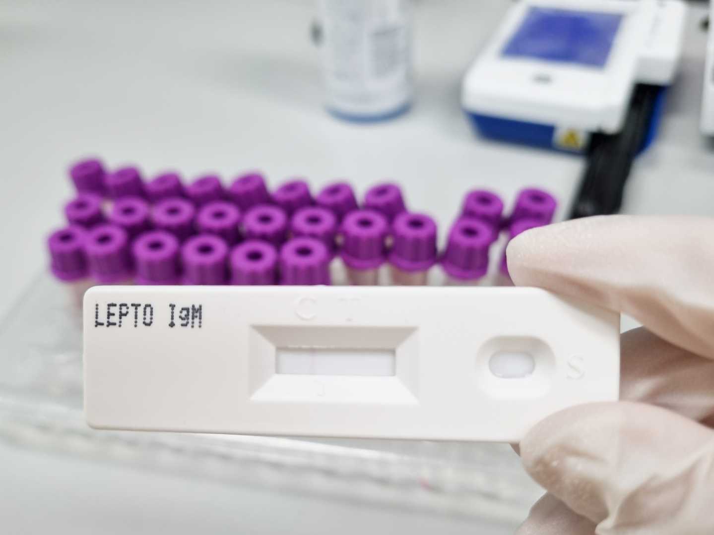
This is the gold - standard serological test and the most widely used diagnostic test. It is based on the ability of serial dilutions of patient serum to agglutinate live leptospiral serovars in vitro. However, MAT has many shortcomings. It is complex to control, perform, and interpret. It is time - consuming, requires the maintenance of live cultures of several pathogenic serovars, and needs special equipment and technical expertise. Results can be subjective and prone to inter - laboratory and intra - laboratory variation.
Enzyme - linked Immunosorbent Assay (ELISA)
ELISA stands as the most common technique for identifying anti - leptospiral antibodies. The method provides multiple advantages which include fast processing time and user-friendly operation while ensuring safety and the ability to analyze numerous samples effectively. The ELISA technique enables simultaneous detection of IgM and IgG antibodies. The IgM ELISA system identifies antibodies 4 to 5 days following symptom onset which is sooner than MAT detection capabilities. The IgG ELISA method helps measure protective immunity duration.
Creative Diagnostics provide high-quality Leptospira specific ELISA kits, featuring high sensitivity and specificity, enabling accurate detection of leptospiral antibodies in serum samples.
Leptospira Hardjo Antibody ELISA Kit (DEIA2332)
PCR

PCR has been increasingly used for the diagnosis of leptospirosis in recent years and tends to replace serological methods in endemic zones due to its sensitivity and capacity for early diagnosis. Real - time PCR (SYBR Green or Taqman technology) is faster than regular PCR and less sensitive to contamination. Several regular or real - time PCR methods have been developed for the detection of pathogenic leptospires, but only a few have been clinically validated. A positive PCR reveals the presence of pathogenic leptospires in the sample but does not directly identify the serovar.
Isothermal Amplification Methods
These methods, such as Loop - mediated isothermal amplification (LAMP), require just a heating unit to maintain a constant temperature of 60 to 65 °C, making them suitable for developing countries. An effective and specific amplification can be performed in 1 hour with polymerase DNA and six primers. The amplified DNA can be detected by simple eye - observation of fluorescence or turbidity without using electrophoresis gels.
References
Loading ......
