
Inquiry Basket
 Loading ......
Phage display technology functions as a screening method which stems from genetic engineering and molecular biology principles and stands out because of its high throughput capabilities alongside cost-effective efficiency. The fundamental mechanism of phage display technology involves combining the gene that encodes an exogenous peptide or protein with the gene for the phage coat protein so that the target molecule appears on the phage particle surface but the phage remains infectious. The technology provides a direct connection between genotype and phenotype which permits high-throughput screening together with functional analysis of target molecules. Smith introduced the concept of utilizing genetic engineering to merge combinatorial peptide libraries with phages for phage display creation in 1985. The researcher determined that exogenous proteins displayed on the phage surface maintain structures and functions comparable to natural proteins. Following this discovery researchers proposed and validated the use of random peptide libraries for antigen epitope identification which triggered the extensive development of phage display technology.
Phage display technology involves inserting an exogenous gene segment into an appropriate site of the phage structural gene using genetic engineering. When the phage infects Escherichia coli, the exogenous gene and the structural gene are expressed as a fusion protein. The displayed peptide or protein retains good biological activity and preserves the phage's original physiological functions and viability. The phage library is then subjected to biopanning to wash away free phages that do not bind the target, selecting phages that specifically bind to the target molecule. The specifically bound phages are eluted using acid and amplified by infecting host cells. Typically, 3–5 rounds of "binding–elution–amplification" are performed to highly enrich phages that specifically bind the target molecule. The selected phages are sequenced and analyzed bioinformatically to obtain information about the target peptide, which is then chemically synthesized for further study.
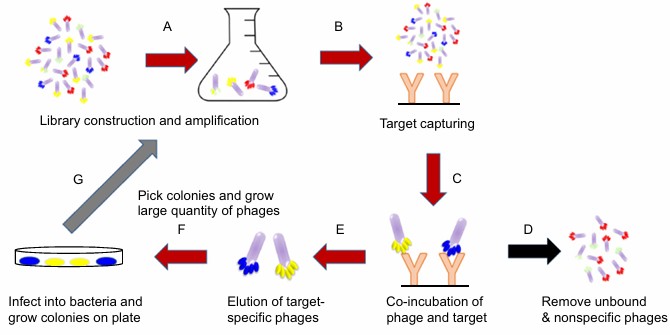 Figure 1. The general scheme of phage display technique and biopanning selection
(Source: Saw PE, et al. 2019)
Phage display systems mainly rely on filamentous phages such as M13, fd, and f1, with their coat proteins pIII and pVIII serving as the primary sites for fusion and display of exogenous molecules. pIII is the largest coat protein, containing multiple repeats of the -SGG- or -SGGG- sequence, and its spatial structure is flexible and variable. The C-terminus of pIII is hydrophobic and anchors to the phage coat, while the N-terminus is free and exposed outside, allowing large fragments of foreign proteins to be inserted at the N-terminus without affecting the phage's structure or function. The pVIII protein is the main component of the phage coat, with about 2,700 copies per phage, making it suitable for displaying short peptides. The C-terminus of pVIII mainly contains basic amino acid residues that easily bind DNA; the middle region is hydrophobic, forming the phage coat; and the N-terminus is free and exposed on the phage surface. Fusion proteins with pVIII allow multivalent display, which can be used for affinity-based screening of target genes. By inserting foreign genes into specific sites of the coat protein genes, the phage replicates and assembles in the host Escherichia coli, co-expressing the foreign proteins with the coat proteins and displaying them on the phage surface.
 Figure 2. Schematic representation of the M13 bacteriophage
(Source: Ledsgaard L, et al. 2018)
In addition, other phage display systems have also been used as vector systems. The λ phage display system fuses exogenous peptides or proteins by inserting them at the N- or C-terminus of the λ phage head assembly protein gpD, or into the carboxyl-terminal folding region of the major tail protein gpV. The λ phage does not need to secrete the exogenous peptides or proteins outside the bacterial cell membrane; assembly can be completed directly inside the host cell. This system has a wide range of applications, capable of displaying active large proteins as well as proteins toxic to host cells. However, it is less suitable for screening high-affinity ligands and has certain limitations. The T4 phage display system displays exogenous peptides or proteins by fusing them to the C-terminus of the SOC or HOC sites. Display at the SOC site is achieved by using an integrative vector to homologously integrate the SOC fusion gene containing the exogenous sequence into a T4 genome lacking SOC, selecting phages that grow independently of lysozyme to achieve display. Display at the HOC site is done through in vitro packaging: the target gene is inserted at the C-terminus of the HOC gene to construct an HOC fusion expression vector. The expressed HOC fusion protein is packaged onto the HOC site on the T4 phage capsid surface when infecting a T4 mutant lacking the HOC protein, thus achieving display. The T4 phage display system has a large capacity and can be assembled in vitro, but since it uses C-terminal fusion, it is not suitable for studies involving the N-terminus of proteins, limiting its applications.
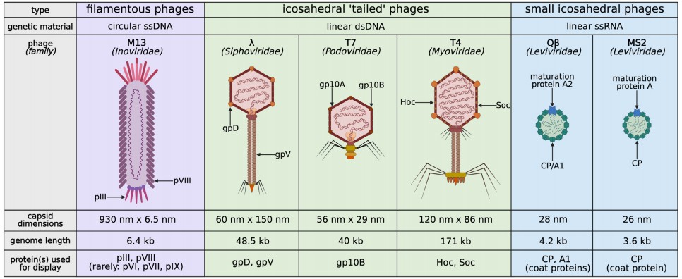 Figure 3. Types of bacteriophages used in phage display
(Source: Jaroszewicz W, et al. 2022)
Scientists use various methods to display antigenic epitopes of different pathogens on phage surface particles for immunization in mice. In this research, they found that phage display libraries, in which each viral particle expresses a different protein or peptide, have broader applications. These libraries include antibody libraries, cDNA libraries, RNA libraries, peptide libraries, and even protein libraries. Compared to other types of expression vectors, phage display libraries have several advantages. First, since the DNA encoding the displayed peptide or protein is packaged within the same particle, there is a physical link between the DNA and the expressed peptide or protein, making it easy to identify interacting peptides through DNA isolation, amino acid sequencing, and bioinformatics analysis. Second, phage particles are very small; a 1 mL solution may contain 1012 phage particles, allowing high-throughput screening with different phage particles in each round. The library screening process is called "panning," which enables relatively easy identification of a specific phage.
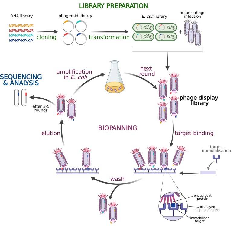 Figure 4. Library preparation and biopanning procedures based on phagemid and helper phage M13 pIII display
(Source: Jaroszewicz W, et al. 2022)
A phage display random peptide library is a molecular library constructed by displaying peptides with various local three-dimensional structures, composed of different arrangements of amino acids, on the surface of the phage. Phage display random peptide libraries can screen linear and mimetic epitopes and can link protein function with genetic information to study protein-protein interactions, such as finding binding sites for ligand-receptor interactions and exploring high-affinity ligand molecules. Because random peptide libraries can recognize self-antigens, this technology can be used to screen and identify antigenic epitopes that specifically bind to autoantibodies associated with autoimmune diseases. A phage display antibody library is a phage library formed by expressing the light and heavy chain gene repertoire of a complete set of antibodies amplified from B lymphocytes through a phage surface display system. Natural phage antibody libraries use B lymphocytes isolated from human or animal blood, bone marrow, spleen, and other tissues or organs, extract RNA, and use synthesized heavy and light chain primers to amplify antibody variable region genes (VH and VL) by RT-PCR. PCR is then used to construct VH and VL into single-chain antibodies (scFv) and clone them into phage expression vectors, and the antibody genes are displayed on the phage surface using helper phages, resulting in a natural phage antibody library. These antibody libraries were the first to be reported as successfully used in monoclonal antibody preparation. Synthetic antibody libraries are divided into semi-synthetic and fully synthetic antibody libraries. Semi-synthetic antibody libraries are based on the framework regions and CDR1 and CDR2 regions of natural antibody libraries, with the DNA sequence of the CDR3 region synthesized randomly and amplified to obtain a semi-synthetic antibody library. Fully synthetic antibody libraries are purely artificial antibody libraries designed and synthesized based on antibody gene information. The variable region genes (VH and VL) of the light and heavy chains used to construct the antibody library are derived from synthetic gene fragments.
Phage display technology can effectively target and identify tumors and organs. Scientists detected specific serum biomarkers for cystic fibrosis by immunoscreening T7 phage display libraries. Doctors can identify cystic fibrosis which affects the CFTR protein by measuring specific biomarkers through phage display technology which also helps create molecular treatments. Phage-based bioassays show more promise for future advancement than conventional antibody-based immunoassays. Shorter phages are easier to bind or genetically modify. In recent years, phage biosensors have advanced significantly due to their good stability, sensitivity, and selective binding. Researchers have developed biosensors for cardiac troponin I using peptides obtained from phage display; peptides screened from phage display libraries serve as sensing probes in these biosensors. Compared to other complex affinity scaffolds, short peptides have several advantages in biosensing applications: 1) peptide synthesis is simple and inexpensive; 2) peptides have more stable bioactivity and better tolerance to harsh environments; 3) peptides are easier to control than engineered antibodies at the molecular level.
Phage display peptide libraries have strong advantages in vaccine development research, bringing new hope for disease prevention and treatment. Phage-based vaccine immunogenicity depends on selecting B-cell epitopes (BCEs) and T-cell epitopes (TCEs). Phage display vaccines enter host cells through endocytosis and antigen-presenting cells process and present them to activate CD4+ and CD8+ T cells by MHC class I and II pathways while simultaneously triggering humoral responses through B cell presentation. Research demonstrates fd filamentous phages activate the complete spectrum of immune responses particularly through surface-displayed MHC class I-restricted epitopes which induce cytotoxic T lymphocyte (CTL) responses. Other research has found that fd phages can target mouse dendritic cells (DCs) and activate innate and adaptive immune responses without the need for exogenous adjuvants.
PALT is an antibody production technology based on phage display technology (PDT). It does not rely on in vivo immune responses and can be used to discover antibodies against almost any type of antigen. Combining the unified features of PDT expressing both protein genotype and phenotype, antibodies corresponding to target genes can be obtained through phenotypic screening, which is useful for identifying disease biomarkers and screening antibody drugs.
The basic construction process of PALT is as follows: Various cells cultured in l undergo total RNA extraction including immune cells together with hybridoma cells and B lymphocytes from peripheral blood bone marrow and spleen. Researchers generate a cDNA library from total RNA through reverse-transcription followed by amplification of the full set of variable region genes including VH and VL genes. VH and VL genes are randomly combined to construct a diverse antibody gene library. The antibody genes are cloned into phage expression vectors, and helper phages are added to infect Escherichia coli. After centrifugation, the supernatant contains the primary phage antibody library targeting a specific antigen. A specific antigen is then selected as the target, and the primary antibody library undergoes 3 to 6 rounds of repeated screening.
The screening process mainly involves two steps: panning and monoclonal identification. Panning consists of adsorption, elution, and amplification. Adsorption refers to incubating the phage antibody library with the antigen to allow specific binding between phage antibodies and the antigen. Unbound phages are discarded, and phages specifically bound to the antigen are eluted without affecting their infectivity and collected. The eluted phages are then used to infect bacteria and amplified for the next round of panning. After several rounds of panning, the enriched polyclonal phages specifically binding the antigen are monoclonalized by selecting single clones from the polyclonal phage population. This yields highly specific monoclonal strains and obtains the corresponding antibody genes.
References
| Target | Cat. No. | Product Name | Size | Species | Application | Detection Sample | |
| Phage | DEIA592 | Phage ELISA Kit | 96T | Quantitative | Specimen containing M13 | Inquiry |
| Target | Cat. No. | Product Name | Expression System | Tag/Conjugate | Application | |
| cre | DAG-P2072 | Bacteriophage P1 cre peptide | N/A | Unconjugated | ELISA | Inquiry |
| Acyrthosiphon pisum secondary endosymbiont phage 1 p50 | DAGA-3445 | Recombinant Protein Acyrthosiphon pisum secondary endosymbiont phage 1 p50, Mammalian cell | Mammalian cell | His | N/A | Inquiry |
| Acyrthosiphon pisum secondary endosymbiont phage 1 p41 | DAGA-3446 | Recombinant Protein Acyrthosiphon pisum secondary endosymbiont phage 1 p41, Baculovirus | Baculovirus | His | N/A | Inquiry |
| Acyrthosiphon pisum secondary endosymbiont phage 1 p31 | DAGA-3447 | Recombinant Protein Acyrthosiphon pisum secondary endosymbiont phage 1 p31, E.coli | E. coli | His | N/A | Inquiry |
Loading ......
