
Inquiry Basket
 Loading ......
Mycobacterium tuberculosis (MTB) represents a Gram-positive bacterium that is acid-fast and requires oxygen to survive as an obligate aerobic bacillus. The organism shows a long rod-shaped structure which can occasionally display slight curves and lacks any flagella or spores. Polysaccharides and lipids with distinctive and unusual structures form the primary composition of MTB cell walls. During infection Mycobacterium tuberculosis depends on its cell wall components to shield against environmental factors and control the host immune response which makes these components vital in tuberculosis pathogenesis. The MTB cell wall's lipid components determine its pathogenicity because increased lipid content strengthens its virulence.
The MTB cell wall, from inside to outside, consists of the cell membrane, peptidoglycan layer, outer membrane layer, and capsule. PG is covalently linked to arabinogalactan (AG), which is esterified at its non-reducing end with α-alkyl, β-hydroxy long-chain mycolic acids. The outer membrane is an asymmetric bilayer mainly composed of MA and phospholipids, with non-covalently embedded lipids and lipopolysaccharides such as phosphatidylinositol mannoside, phthiocerol dimycocerosate, phenolic glycolipid, mannose-capped lipoarabinomannan (ManLAM), and various acyl trehaloses. ManLAM is anchored to the bacterial cell membrane through the PIM anchor and extends through the cell wall to the outer membrane surface. The outermost layer is a loosely attached capsule mainly composed of α-D-glucan, proteins, and a small amount of lipids. The capsule also contains D-arabino-D-mannan.
 Figure 1. M. tuberculosis cell wall
(Source: Jacobo-Delgado YM, et al. 2023)
The cell wall antigen LAM plays an essential role in mycobacteria by modulating immune responses during interactions with hosts. The LAM capping process produces ManLAM in slow-growing pathogenic species including MTB and Mycobacterium bovis and Mycobacterium leprae but results in PILAM in fast-growing non-pathogenic species like Mycobacterium smegmatis. ManLAM's mannose cap functions to inhibit pro-inflammatory responses while PILAM's phosphatidylinositol cap triggers human macrophages to release pro-inflammatory cytokines. Mycobacterium phlei is an example of other fast-growing mycobacteria that produce LAM molecules without mannose or phosphatidylinositol caps called AraLAM.
LAM is one of the three major interrelated lipoglycan groups in the mycobacterial cell wall. The LAM molecule is non-covalently anchored to the mycobacterial plasma membrane via a glycolipid anchor and extends outward to the cell wall surface. It consists of three main parts: (1) a glycosylphosphatidylinositol anchor that attaches it to the plasma membrane; (2) a highly conserved core structure that links to the glycolipid anchor found in mycobacteria; and (3) an arabinan domain, which includes variable mannose caps and side chains. Different structural variations generate LAM molecules that exhibit distinct properties and functional roles. Mannose caps enable mycobacteria to attach to macrophage mannose receptors which creates an ideal intracellular setting for the pathogen.
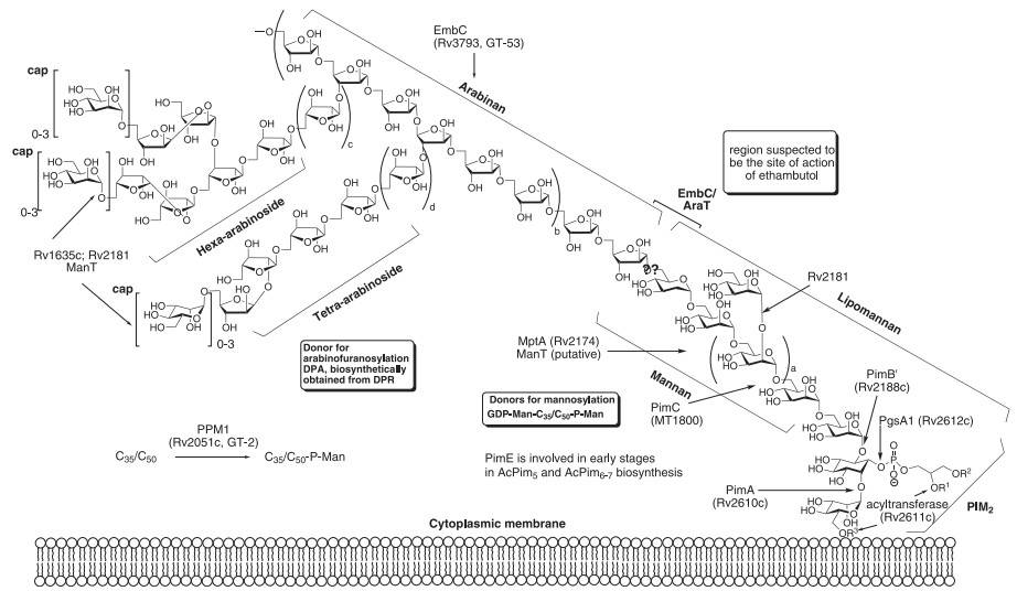 Figure 2. Current model of M. tuberculosis ManLAM with structurally related components PIMs, LM, and LAM
(Source: Umesiri FE, et al. 2010)
The LAM structure from highly virulent Mycobacterium tuberculosis strains such as H37Rv, Erdman, or NYH-27 exhibits structural differences when compared to the LAM of both avirulent strains and clinical isolates as well as other pathogenic mycobacteria including Mycobacterium leprae. The structural differences and diversity of LAM are mainly determined by: Terminal linked molecules like mannose, arabinose, or methylthio-D-xylose (MTX) determine complex structural components while the epitope structural configuration together with the MTX branch linked to Man establish epitope length and composition. It has been observed that these structural components of LAM can change dynamically, affecting epitope recognition. Changes in LAM structure directly influence the specificity of some antibody epitope recognition. LAM is unstable as a monomer in blood and binds to carrier molecules like high-density lipoprotein in serum. It circulates through the blood and passes through the kidneys. Because LAM is antigenic, it cannot pass directly through the glomerulus but is filtered into urine as immune complexes, resulting in higher LAM concentration in urine than in serum.
Mycobacterium tuberculosis has strong adaptability under stress conditions and can undergo proteome and lipidome rearrangements. During different stages of infection, the distribution of glycolipids and the ratio of LAM to lipomannan (LM) can change. Low concentrations of LAM can induce negative feedback regulation of the immune system, possibly because LAM has multiple epitopes that interact with cell surface receptors with varying affinities. For example, the lectin domain of CR3 exposed on eosinophils can interact with mannose, mediating bacterial uptake. The variety of mannose units in LAM influences dendritic cell maturation and function as their concentration increases. LAM extracted from Mycobacterium tuberculosis and Mycobacterium leprae functions by suppressing T cell multiplication while also interfering with interferon-gamma-driven macrophage activation and protein kinase activity and by triggering tumor necrosis factor production in monocytes and complement activation.
The virulence factors of MTB such as LAM modulate innate immune responses by altering the functions of immune cells including alveolar epithelial cells, macrophages and dendritic cells. The production of IL-37 in human type II alveolar epithelial cells is triggered by LAM through TLR-2 initiated signaling pathways which result in p38 upregulation and ERK1/2 phosphorylation. IL-37 binds to Smad3 and moves into the nucleus to block pro-inflammatory cytokine signals. LAM can block activation and differentiation of immune cell subsets. In vitro, monocytes exposed to LAM fail to differentiate into mature macrophages, showing reduced expression of CD86, TLR2, and TLR4. These macrophages have impaired tumor necrosis factor (TNF) signaling and defective PAR2 pathway function, weakening control of intracellular MTB growth. LAM targets neutrophils through insertion into lactosylceramide-enriched lipid rafts on their membranes which disrupts tyrosine kinase signaling and blocks hematopoietic cell kinase recruitment while reducing granule trafficking. The production of myeloperoxidase and NADPH oxidase decreases while calcium flux becomes impaired which restricts phagolysosome formation and cytokine production as well as degranulation and respiratory burst. LAM binds to CD11b/CD18 receptors on eosinophils which stops TLR-2 activation and restricts the release of inflammatory cytokines. LAM inhibits phagosome maturation, further impairing innate immune activation. Genetic polymorphisms such as TLR1 1805G/T are associated with increased susceptibility to MTB due to altered LAM recognition. Tuberculosis patients have circulating LAM which attaches to high-density lipoprotein to enhance macrophage vulnerability to mycobacteria while reducing TNF production substantially. The presence of LAM modifies innate immune cells in a way that slows down their response to MTB infection and reduces their effectiveness.
 Figure 3. Schematic illustration of MTB-host interactions with and inside of the alveolar macrophage
(Source: Naeem MA, et al. 2021)
In regulating adaptive immunity, LAM promotes an increase in the number of IL-10-producing B cell subsets. An IL-10-rich environment allows Mycobacterium tuberculosis to survive by suppressing the host's pro-inflammatory immune defenses. LAM suppresses T cell activation through its effect on reducing antigen-presenting molecule expression including MHC-II and CD80/CD86. When dendritic cells undergo treatment with LAM they display diminished ability to cross-present tuberculosis antigens which leads to CD4+ T cell proliferation dropping by 40% to 60%.
An ideal diagnostic technique for tuberculosis should distinguish between active tuberculosis patients, latent tuberculosis infection individuals, and cured cases. LAM, released from the MTB cell wall or present on its surface, serves as a unique biomarker in tuberculosis diagnosis. It has potential value in differentiating LTBI from active tuberculosis and is diagnostically valuable for HIV-positive tuberculosis patients and children who cannot produce sputum. LAM detection is relatively low-cost, and ManLAM in sputum can serve as a biomarker for bacterial load before and after tuberculosis treatment, helping to evaluate treatment efficacy.
To further improve the detection performance of LAM, research on anti-LAM monoclonal antibodies has been ongoing. Currently, the prepared monoclonal antibodies mainly include those that detect the Ara4 and Ara6 structures of LAM, as well as antibodies strictly dependent on the Ara6 structure. These include natural human monoclonal antibodies, mouse-derived monoclonal antibodies, and four types of monoclonal antibodies derived from phage display libraries.
Research demonstrates that glomerular endothelial cells create a porous mesh structure during kidney blood filtration which permits MTB-bound LAM to traverse membrane molecules or extracellular vesicles and enter the urine for excretion. An ELISA detection method can measure LAM levels in urine for tuberculosis diagnosis. The urine LAM detection method demonstrates high sensitivity when identifying tuberculosis in HIV patients who experience advanced immunodeficiency and possess low CD4 cell counts. LF-LAM detection represents a commercial bedside test for active pulmonary tuberculosis and receives WHO endorsement as one of the recommended bedside tests for tuberculosis. This test helps diagnose tuberculosis in severely ill HIV patients who have low CD4 cell counts. The main advantage of LF-LAM is its low cost, but its sensitivity is poor, and LAM testing is not recommended for patients with CD4 counts above 200 cells/mm³. The urine LAM test demonstrates multiple limitations which include reduced detection sensitivity in tuberculosis-exclusive patients, poor monoclonal antibody specificity, and diagnostic analysis interference due to tuberculosis patients' clinical characteristics.
Since the discovery that LAM can bind to cell membranes and exhibits amphipathic properties, new approaches have emerged for immunoassay detection of Mycobacterium tuberculosis. This method can detect LAM not only in histological specimens but also in peripheral blood. ELISA detection of LAM antigen in blood showed a sensitivity of 88% for smear-positive active pulmonary tuberculosis patients, 67% for smear-negative active pulmonary tuberculosis patients, and only 57% for HIV-tuberculosis co-infected patients. The specificity for tuberculosis patients was 91.5%. Compared to urine LAM detection, blood LAM-ELISA has lower sensitivity in immunosuppressed tuberculosis patients. LAM can move within bacterial vesicles, which may help it evade antibody recognition. Serum LAM detection has limitations such as low sensitivity for tuberculosis-only patients, low monoclonal antibody specificity, and interference from clinical features of tuberculosis patients in diagnostic analysis.
References
| Target | Cat. No. | Product Name | Host | Isotype | Application | |
| LAM | CABT-YN1556 | Mouse Anti-M. tuberculosis TB-LAM Monoclonal Antibody, clone 5D17 | Mouse | IgG | ELISA (Cap), CLIA, LFIA | Inquiry |
| CABT-YN1557 | Mouse Anti-M. tuberculosis TB-LAM Monoclonal Antibody, clone 3F2 | Mouse | IgG | ELISA (Det), CLIA, LFIA | Inquiry | |
| CABT-NS1189 | Rabbit Anti-LAM Monoclonal Antibody, clone CS-35 | Rabbit | IgG | ELISA, WB, FC, IF | Inquiry | |
| CABT-BL2113 | Anti-Lam monoclonal antibody, clone ADL46 | Mouse | IgG1 | WB | Inquiry | |
| CABT-L0374Y | Humanized anti-M. tuberculosis lipoarabinomannan(LAM) monoclonal antibody, clone 2B5 | Human | IgG | ELISA, CLIA, ELISA(Cap), CLIA(Cap) | Inquiry | |
| CABT-L0375Y | Humanized anti-M. tuberculosis lipoarabinomannan(LAM) monoclonal antibody, clone 6D4 | Human | IgG | ELISA, CLIA, ELISA(Det), CLIA(Det) | Inquiry | |
| M. tuberculosis LAM | DMAB-CS23046 | Rabbit Anti-M. Tuberculosis LAM Monoclonal antibody, clone 2B5 | Rabbit | IgG, kappa | ELISA, IF, LFIA | Inquiry |
| Target | Cat. No. | Product Name | Expression System | Tag/Conjugate | Application | |
| M. tuberculosis LAM | DAGA-168 | Native M. Tuberculosis LAM antigen | Mycobacterium tuberculosis | N/A | ELISA, CLIA, LF | Inquiry |
Loading ......
