
Inquiry Basket
 Loading ......
Glycosphingolipids (GSLs) are a class of amphipathic lipids with ceramide as the parent structure, which can be classified as acidic, basic, and neutral glycosphingolipids, and play an important role in life activities such as cell growth, apposition, cell cycle, cell differentiation, and tumorigenesis. GSLs have been shown to be involved in a variety of immune processes, including differentiation, recognition, recruitment of proteins to specific membrane microdomains, direct interaction with surface receptors, and transduction of activation signals.
Globotriaosylceramide (Gb3) is a neutral glycosphingolipid found in the detergent-insoluble portion of the membranes of cholesterol-rich lipid rafts, and is particularly abundant in the cell membranes of CNS cells, renal endothelial cells, intestinal epithelial cells, and some tumor cells, etc., where it plays a very important role in cell growth, differentiation, recognition, and signal transduction. During the discovery of Gb3, it was given various names, depending on the field of study and the cells chosen at the time. Those found in erythrocytes were called Pk blood group antigens, those found in lymphocyte studies were called CD77 differentiation antigens, and those found in some endothelial cell studies were called Shiga toxin (STx) receptors. CD77 is expressed predominantly on B cells and plays a key role in B cell development, antibody production, and in tumors and autoimmune diseases. Gb3 is a natural receptor for STx and, by binding to Shiga Toxin B subunit (STxB), assists in the internalization of the toxin thereby exerting its toxic effects and causing a range of intestinal disorders. In addition, a disorder of glycosphingolipid catabolism known as Fabry disease leads to intracellular deposition of Gb3 in the vascular endothelium and other tissues. This inherited disease is a heterogeneous progressive disorder with manifestations including numbness of the extremities, sweating abnormalities, angiokeratomas, and cardiovascular, cerebrovascular, and renal disease due to dysfunction of the organs affected by Gb3 accumulation. Gb3 is also another cofactor for CD4-dependent HIV-1 fusion, preferentially interacting with CXCR4 gp120.
Gb3 is formed by a lipid backbone connecting the oligosaccharides, and the lipid portion consists of a sphingosine and a fatty acid bonded via an amide bond, resulting in a ceramide molecule. Gb3 molecules in which the sphingosine portion is modified are usually referred to as analogues, and Gb3 molecules in which the fatty acid chain is modified are referred to as isoforms. Fatty acids and sphingosines vary in length from 16 to 26 carbon atoms. The different Gb3 analogues and isomers can be divided into five groups based on chemical modifications. Group 1 consists of Gb3 isomers with saturated fatty acids and Group 2 consists of Gb3 isoforms/analogues with an additional double bond (in sphingosine or fatty acids). Group 3 are Gb3 isoforms/analogues with two additional double bonds, Group 4 are hydroxylated fatty acid Gb3 isomers and Group 5 are methylated Gb3 isomers. Notably, these modifications alter the affinity of Stx for Gb3 binding.
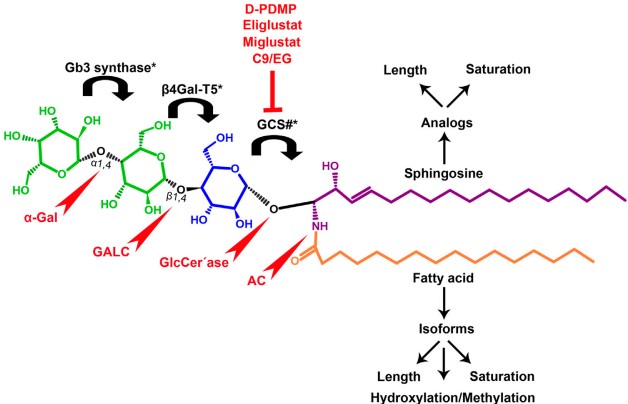 Figure 1. Chemical structure of Gb3
(Source: Celi AB, et al. 2022)
Ceramide molecules are synthesized from sphingosine and fatty acids in the cytoplasm of the endoplasmic reticulum membrane by ceramide synthase (CerS1-6). The oligosaccharide portion of Gb3 is synthesized in the Golgi. Firstly glucosylceramide synthase (GCS) catalyses the binding of a glucose molecule to ceramide to synthesize glucosylceramide (GlcCer), followed by β1,4 galactosyltransferase 5 (β4Gal-T5) binding a galactose molecule to form lactosylceramide (LacCer). Finally, Gb3 synthase catalyses the addition of α-1,4-galactose to LacCer to produce Gb3. During Gb3 synthesis, the newly formed ceramides are transported through the membrane to reach the cis-Golgi cisternal, where the cytoplasmic enzyme GCS generates GlcCer in the Golgi cytoplasmic leaflet. Gb3 on the cell membrane is in dynamic equilibrium, newly synthesised Gb3 is transported to the cell membrane via the secretory efflux pathway, while Gb3 on the cell membrane can be translocated along the endocytosis pathway via the endosomes to the lysosomes or to the Golgi apparatus and endoplasmic reticulum.
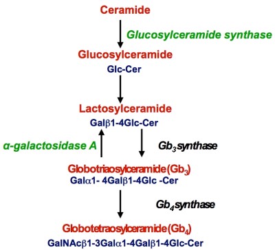 Figure 2. Biosynthesis of Gb3
(Source: Lingwood CA, et al. 2011)
Degradation of Gb3 occurs in lysosomes in four steps in the presence of acid hydrolases. Acid hydrolase successively eliminates each carbohydrate residue in Gb3, a process that ends with the elimination of the fatty acid chains in ceramide, resulting in the production of three free hexoses, one free fatty acid chain, and one sphingosine. Deficiencies in the enzymes responsible for degradation lead to Fabry, Gaucher disease, which are characterized by the accumulation of GSL (including Gb3) in lysosomes in different cell types. C-9 is a specific inhibitor of GCS that reduces the synthesis of Gb3 and therefore can be used as a therapeutic agent for these diseases.
GSL synthesis is regulated by cytokines such as IFN-α, TNF-α, and IL-1. IFN-α increases the expression of Gb3 regulation-related genes, leading to an imbalance in GSL accumulation. TNF-α increases Gb3 synthesis via GCS and upregulates Beta4Gal-T5 and Gb3 via NF-κB signaling in the mitogen-activated protein kinase (p38, JNK 1/2, ERK 1/2)-induced cascade.
Gb3 has long been thought to be specifically expressed by germinal center B cells during apoptosis, and was originally postulated as a mechanism for eliminating cells that fail to produce high-affinity antibodies during antigen-driven selection in secondary lymphoid organ germinal centers. Subsequent studies revealed that Gb3 is expressed in a subpopulation of germinal center B cells involved in somatic mutations of the immunoglobulin (Ig) gene. These cells are known as centroblasts and do not undergo apoptosis. CD77 promotes the activation and proliferation of B cells through interactions with other surface receptors (e.g. CD19, CD20, etc.). This process is essential for antibody production and the formation of memory B cells. In recent years, it has been found that CD77 also plays an important role in the tumor microenvironment. It is not only involved in the immune escape mechanism of tumor cells, but may also affect tumor development and metastasis. Some tumor cells can up-regulate CD77 expression to evade attack by the host immune system. This mechanism may be achieved by inhibiting T cell activation or inducing the production of regulatory T cells (Treg). In addition to its role in tumors, CD77 is associated with a variety of autoimmune diseases. For example, the expression level of CD77 is often abnormal in diseases such as systemic lupus erythematosus (SLE) and rheumatoid arthritis. This suggests that CD77 may play an important role in the development of these diseases. It was found that CD77 expression was upregulated in SLE patients and correlated with disease activity. This suggests that CD77 may be used as a biomarker to assess the disease in SLE patients. In patients with rheumatoid arthritis, CD77 also shows aberrant expression that correlates with arthritic symptoms. This provides a rationale for the development of novel therapeutic strategies targeting CD77.
Gb3 is also important for the FAS receptor-ligand system, which is one of the major mechanisms for the elimination of germ-infected cells and for the control of autoimmune diseases and certain malignancies. The cell surface binding receptor FAS belongs to the TNF receptor family and contains an intracellular death structural domain that triggers apoptosis. The physiological ligand FASL is a member of the corresponding TNF cytokine family and is expressed on the surface of classical CD8+ cytotoxic T cells and natural killer T cells. FAS internalisation is required for the activation of the apoptotic pathway. FAS has a conserved extracellular Gb3-binding motif that interacts strongly with lactosylceramide and weakly with the dibasic gangliosides GD3 and Gb4, thereby regulating its internalisation pathway leading to clathrin-mediated internalisation of FAS and subsequent cell death signaling. Thus, the interaction of Gb3 with the conserved extracellular Gb3-binding motif is critical for the activation of the apoptotic pathway in FAS.
Gb3 is also a natural receptor for STx, which currently provides a means to develop new cancer treatments. The STx complex, consisting of a toxic catalytic A-subunit and a non-toxic receptor-binding B-subunit pentamer, is a potent tool for a variety of therapeutic approaches, including oncology treatment and imaging. Solo recombinantly expressed STxB is a versatile protein for tumor targeting and vaccine design. And each application depends on the presence of its preferential receptor, Gb3, on target cells. STxB binds to Gb3 on the plasma membrane and is internalised via endocytosis and appears in clathrin-coated pits. When clathrin-dependent uptake is blocked, STxB can continue to continue endocytosis via clathrin-independent pathways. STxB induces the formation of the STxB-Gb3 cluster structural domain, imposes negative curvature on the host membrane, and promotes lipid reorganisation in tubular membrane invaginations. The occurrence of these events culminates in the entry of STxB into the cell and retrograde transport from early endosomes to the trans-Golgi network and endoplasmic reticulum.
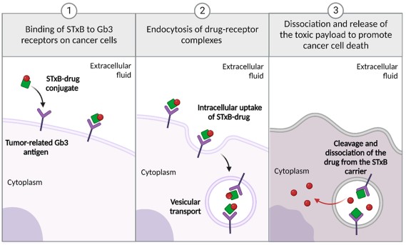 Figure 3. Internalization of Shiga Toxin B subunit enabling a method for drug delivery
(Source: Danielewicz N, et al. 2023)
References
| Target | Cat. No. | Product Name | Expression System | Tag/Conjugate | Application | |
| GB3 | DAG-WT487 | GB3 [BSA] | N/A | BSA | N/A | Inquiry |
| GB3 | DAG-WT488 | GB3 [OVA] | N/A | OVA | N/A | Inquiry |
| Target | Cat. No. | Product Name | Host | Isotype | Application | |
| CD77 | DMABT-Z59126 | Anti-CD77 monoclonal antibody, clone 40-25 | Rat | IgM | FC | Inquiry |
| CD77 | CABT-L1967 | Mouse Anti-Human CD77 monoclonal antibody, clone 6C6 | Mouse | IgM | FC, WB, IHC | Inquiry |
Loading ......
