
Inquiry Basket
 Loading ......
In situ hybridization (ISH) refers to the process of using a specific labeled nucleic acid of known sequence as a probe to hybridize with nucleic acid in a cell or tissue section, so as to accurately and quantitatively locate a specific nucleic acid sequence. ISH can be performed on cell samples or tissue samples and is a direct and convenient method of studying gene localization and expression.
The basic principle of ISH technology is to use the complementary base sequence between the single strands of nucleic acid molecules to complement the exogenous nucleic acid (probe) with the DNA or RNA to be tested on the tissue, cell, or chromosome. The combined specific nucleic acid hybrid molecules can display the position of the nucleic acid to be tested on the tissue, cell, or chromosome through certain detection means.
The types of probes can be divided into two categories: isotope-labeled probes and non-isotope-labeled probes according to the labels they carry. At present, most radioactive labeling methods incorporate labeled genes into DNA through enzymatic reactions. Commonly used isotope labels include 3H, 35S, 125I, and 32P. Although isotopic markers have the advantages of high sensitivity and a clear background, because radioactive isotopes can cause harm to humans and the environment, they tend to be replaced by non-isotopic markers recently. The most commonly used non-isotopic labels include Biotin, digoxin, and fluorescein. Depending on the probe and target nucleic acid used, ISH can be divided into three categories: DNA-DNA hybridization, DNA-RNA hybridization, and RNA-RNA hybridization.
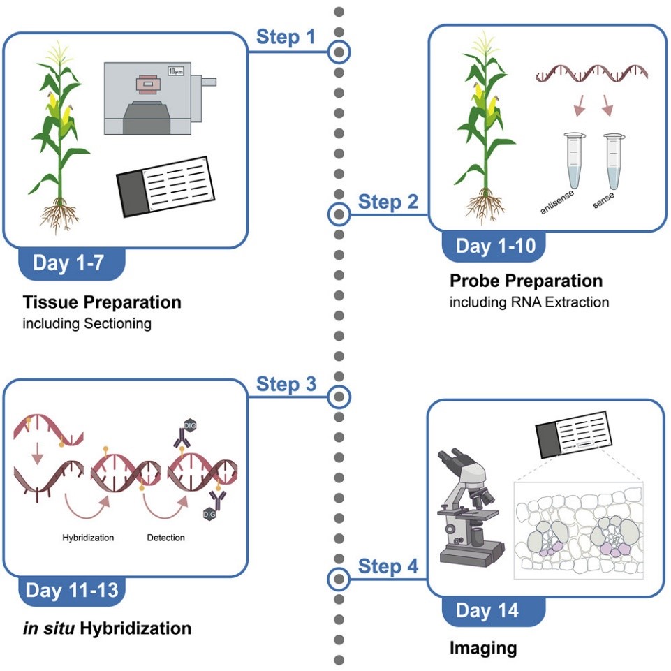 Fig. 1 RNA in situ hybridization protocol for maize leaf tissue. (Zöllner N R,et al., 2021)
The types of probes can be divided into DNA probes, cDNA probes (double-stranded cDNA probes and single-stranded cDNA probes), cRNA probes, and synthetic oligonucleotide probes, depending on the nature of the nucleic acid. Probe selection is critical as it determines the specificity and sensitivity of the assay. The probe must be designed to recognize only the target sequence and not cross-react with other sequences within the sample. If the probe does not precisely match the target sequence, it may only loosely hybridize, leading to potential loss of signal during wash and detection steps and incorrect detection. Therefore, it is essential to ensure that the probe is precisely complementary to the target sequence to achieve optimal results.
When choosing a labeling method, the requirements of the experiment, such as sensitivity and display method, should be considered. When detecting single-copy gene sequences, a probe labeling method with high labeling efficiency and display sensitivity is generally selected. When the requirement for sensitivity is not high, biotin probe technology with a long storage time and a relatively stable alkaline phosphatase display system can be used.
Tissue sampling: Tissue sampling should be as fresh as possible. Because tissue RNA degrades quickly, fresh tissues and cultured cells are best fixed within 30 min.
Cell tissue fixation: The purpose of cell tissue fixation is to maintain cellular structure, maximize the level of DNA or RNA in the cell, and make it easy for probes to enter the cell or tissue.
The most commonly used fixative is paraformaldehyde, which, unlike other aldehyde fixatives, does not extensively cross-link proteins and thus does not interfere with probe penetration into cells or tissues.
Once the probe is labeled, it can be hybridized with the target nucleic acid sequence within the sample. This involves incubating the probe with the sample under specific conditions that promote hybridization. The hybridization step can take several hours to overnight, depending on the probe and target sequence used.
Rinsing with different concentrations and temperatures of saline solution can effectively reduce background staining, and washing with RNase solution can remove non-base-paired RNA from the tissue. It is necessary to observe the principle that the salt solution concentration is from high to low, and the temperature is from low to high.
Note: Do not allow slices to dry.
Different color development treatments can be carried out according to the type of nucleic acid probe label. ISH probes are generally labeled with the hapten Digoxigenin, and the substrate is displayed by means of an indirectly added enzyme (enzyme-labeled anti-Digoxigenin antibody). Commonly used enzymes are horseradish peroxidase (HRP) and alkaline phosphatase (AP).
ISH sections of cells or tissues can be used for semi-quantitative detection after color development, but attention should be paid to strictly controlling the identity of the experimental conditions, including the thickness of the section, the amount of nucleic acid preserved, etc.
Reference
Loading ......
