
Inquiry Basket
 Loading ......
Histones are evolutionarily conserved proteins found in the nucleus of eukaryotic cells. These proteins pack DNA into structures called nucleosomes, fundamental units of chromatin, to fit the DNA molecule into the nucleus. Each of these nucleosomes has two subunits, each comprising the core histones H2A, H2B, H3 and H4, and a linker histone called H1 that acts as a stabilizer.
Histone modifications refer to a range of covalent post-translational modifications (PTMs) that occur primarily on the N-terminal tails of histone proteins. These modifications—such as acetylation, methylation, phosphorylation, and ubiquitination—act as dynamic signals that modulate chromatin structure and function. Through the recruitment of specific effector proteins, histone modifications influence key cellular processes including transcription, DNA repair, replication, and epigenetic inheritance.
Histone Modification Description Rule:
Histone structure + Amino acid name + Amino acid position + Modification type.
Examples:
H3K4ac indicates acetylation of lysine at position 4 on histone H3.
H2AK119ub1 represents monoubiquitination of lysine at position 119 on histone H2A.
A large catalogue of histone modifications has been described, each contributing distinct regulatory effects. Histone acetylation, methylation, and phosphorylation are the most frequent alterations in histone tails, while many other modifications have been detected, such as ubiquitination, lactylation, propionylation, crotonylation, and formylation. The modified amino acid sites on histone tails are shown in Fig. 1.
Acetylation is one of the most extensively studied histone modifications because it is one of the first discovered to affect transcriptional regulation. Acetylation typically occurs on lysine residues of histones, catalyzed by histone acetyltransferases (HATs). Negative charges of added acetyl groups neutralize lysines' positive charges, which results in a weakened histone-DNA interaction, leading to a more relaxed chromatin structure that facilitates the access of transcription factors to genomic sequences, thus increasing gene expression.
Histone methylation mainly occurs on the side chains of lysines and arginines, catalyzed by histone methyltransferases (HMTs). Lysines may be mono-, di- or tri-methylated, whereas arginines may be mono-, symmetrically or asymmetrically di-methylated. Unlike histone acetylation, which causes transcriptional activation through weakening histone-DNA interaction, histone methylation increases the basicity and hydrophobicity of histones and affects DNA-transcription factors interaction, activating or inhibiting gene expression.
Histone phosphorylation takes place on serine (S), threonine (T), and tyrosine (Y), predominantly, but not exclusively, in the N-terminal histone tails. Unlike acetylation and methylation, histone phosphorylation establishes interactions between other histone modifications and serves as a platform for effector proteins, leading to downstream cascade events. Histone phosphorylation can have different effects on gene expression depending on the specific context and the location of the phosphorylation site.
The previously described histone modifications result in relatively small molecular changes to amino-acid side chains. In contrast, ubiquitination results in a much larger covalent modification. Ubiquitin is a 76-amino acid protein highly conserved in eukaryotes that is attached to histone lysines (K) via the sequential action of three enzymes, E1-activating, E2-conjugating and E3-ligating enzymes. All core histone proteins can be ubiquitinated, but H2A and H2B are the most common and the most heavily ubiquitinated proteins in the nucleus. H2A and H2B mainly undergo monoubiquitination, which is a reversible process that alters the histone mass and influences nucleosomal dynamics. Histone polyubiquitination generates an irreversible signal for proteasomal-mediated degradation.
Sumoylation is a modification related to ubiquitylation, and involves the covalent attachment of small ubiquitin-like modifier (SUMO) molecules to histone lysines (K) via the action of E1, E2 and E3 enzymes. Sumoylation has been detected on all four core histones. In mammals, sumoylation mainly occurs on H4, and H4 Sumoylation induces transcriptional repression. Histone sumoylation currently stands as the sole described repressive histone mark in budding yeast owing to the lack of repressive lysine methylation marks like H3K9me and H3K27me.
Histone ADP-ribosylation is the process of adding one or more ADP-ribose moieties to specific amino acids (Lys, Arg, Glu, Asp, and Ser) on histone protein by ADP-ribosyl transferases. ADP-ribosylation reactions can be divided into four groups: mono-ADP-ribosylation, poly-ADP-ribosylation, ADP-ribose cyclization, and formation of O-acetyl-ADP-ribose. Notably, these modifications significantly increase upon DNA damage implicating the pathway in the DNA damage response. Histone mono-ADP-ribosylation is performed by the mono-ADP-ribosyltransferases and has been detected on all four core histones, as well as on the linker histone H1.
In addition to the well-known histone modifications mentioned above, there are many other types of histone modifications. In recent years, non-acetyl histone lysine acylations, such as crotonylation, butyrylation and propionylation, have been identified and always share the same "readers", "writers", and "erasers". Other rare histone modifications, including isomerization, biotinylation and citrullination, are reported to be capable of influencing histone methylation. However, the role of these modifications is still not well-established in cancer.
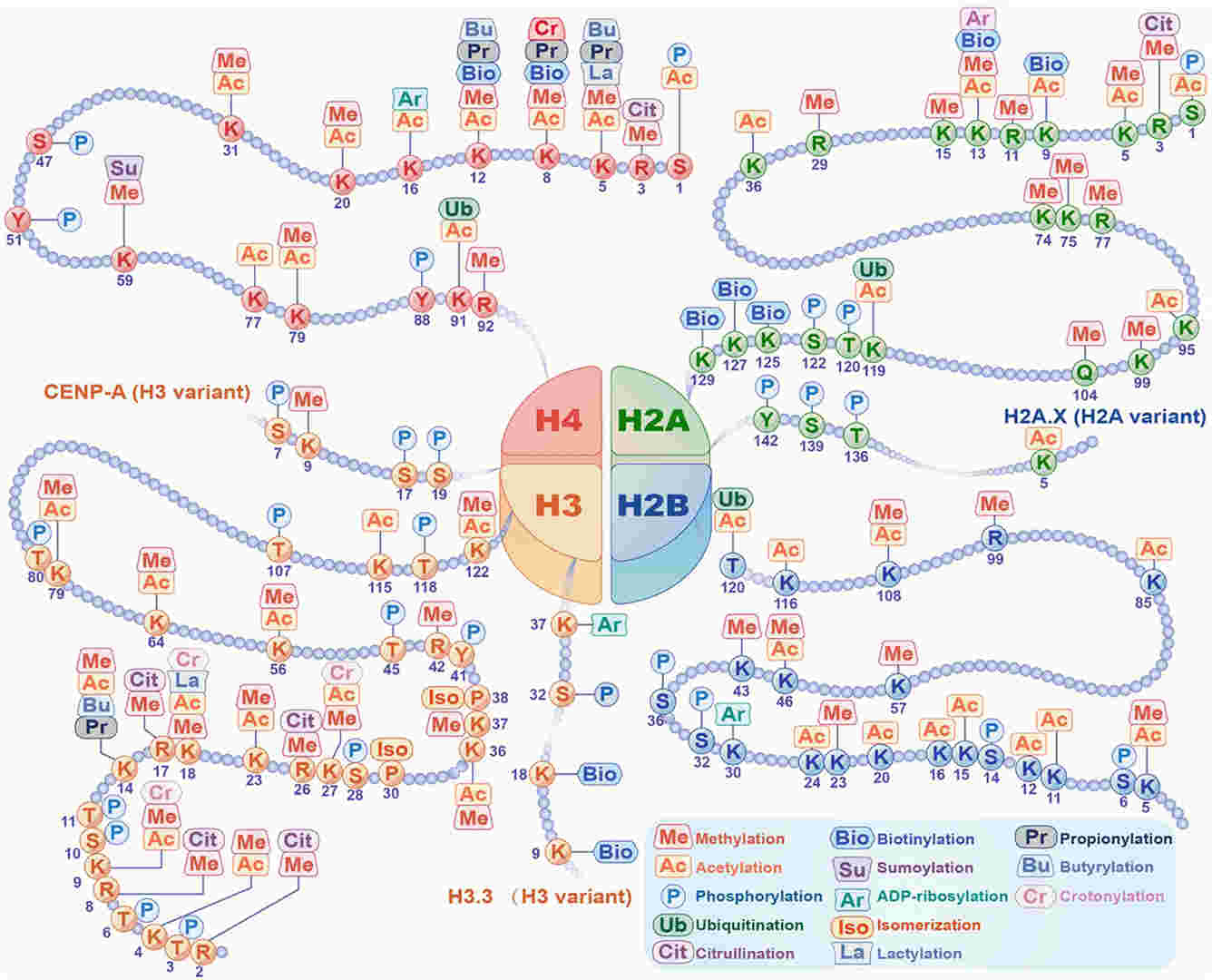 Fig. 1 Histone modifications
Histone modifications are dynamic chemical changes to histone proteins that regulate gene expression. These modifications are controlled by three main classes of proteins:
Writers: These are enzymes that catalyze the addition of chemical groups to histones
Erasers: These are enzymes that remove modifications to alter chromatin states
Readers: These are proteins that recognize and bind specific modifications to mediate downstream effects.
The main histone modifications and their respective writer, eraser enzymes and readers are detailed in the table below.
| Modification | Writers (Adding enzymes) | Erasers (Removing enzymes) | Readers (Recognizing domains/proteins) |
| Acetylation (Ac) | HATs (Histone Acetyltransferases) - CBP/p300 - GCN5 - PCAF - TIP60 | HDACs (Histone Deacetylases) - HDAC1-11 - Sirtuins (SIRT1-7) | Bromodomains - BRD4 - BRD2 - TAF1 |
| Methylation (Me) | HMTs (Histone Methyltransferases) - SET1/MLL (H3K4) - SUV39H1 (H3K9) - EZH2 (H3K27) - SETD2 (H3K36) | HDMs (Histone Demethylases) - LSD1 (H3K4me1/2) - JMJD2 (KDM4 family) - UTX, JMJD3 (H3K27me3) | Chromodomains – HP1 (H3K9me3), Polycomb (H3K27me3) PHD fingers – TAF3 (H3K4me3) Tudor domains – 53BP1 (H4K20me2) MBT domains – L3MBTL1 |
| Phosphorylation (Ph) | Kinases - MSK1/2 (H3S10) - Aurora B (H3S28) - ATM/ATR (H2AX S139, aka γH2AX) | Phosphatases - PP1, PP2A - Wip1 | 14-3-3 proteins – Bind phosphorylated H3 BRCT domains – MDC1 (binds γH2AX) |
| Ubiquitination (Ub) | E3 Ligases - RNF20/40 (H2BK120ub) - RING1B (H2AK119ub) | Deubiquitinases (DUBs) - USP22 - BAP1 - MYSM1 | UIM, UBA domains - RAP80 (DNA repair, binds H2A-Ub) |
| Sumoylation (SUMO) | SUMO E3 Ligases - PIAS1-4 | SENPs (Sentrin/SUMO-specific proteases) - SENP1–7 | SIM (SUMO-Interacting Motif)-containing proteins |
| ADP-ribosylation (ADPr) | PARPs (Poly-ADP-ribose Polymerases) - PARP1, PARP2 | PARG (poly-ADP-ribose glycohydrolase) - ARH3 | Macrodomains, WWE domains, PBZ (PAR-binding zinc finger) – e.g., APLF, CHFR |
| Citrullination (Cit) | PADI enzymes (Peptidylarginine deiminases) - PADI4 (acts on H3R2, H3R8, H4R3) | Generally irreversible (Arg → Cit) | No classical reader domain; affects chromatin by charge change |
Histone modifications serve as fundamental signals that guide a variety of biological processes. Here's a breakdown of the key biological functions:
Transcriptional regulation: Open or compact chromatin to permit or restrict transcription factor access.
Chromatin remodeling: Establish euchromatin (a more open, relaxed state that allows for gene transcription ) or heterochromatin (a more compact, condensed state that silences genes) environments.
Cell cycle and DNA repair: Coordinate genome maintenance and stability.
Cell differentiation and development: Maintain lineage-specific gene expression profiles.
Epigenetic memory: Support stable inheritance of gene expression states across cell generations.
A variety of methods have been developed to detect histone modifications, each tailored to specific experimental objectives such as localization, quantification, or profiling of epigenetic marks. Below are commonly used techniques along with their primary applications:
ChIP is a widely used technique to study the interactions between specific proteins—such as transcription factors or histone modifications—and chromatin. By using antibodies that specifically recognize a particular histone modification, ChIP enriches nucleosomes carrying that modification along with their associated genomic DNA. This allows for precise localization of histone modifications within the genome. The recovered DNA can be analyzed by quantitative PCR (ChIP-qPCR) to assess enrichment at selected loci, or by high-throughput sequencing (ChIP-seq) to generate genome-wide maps of regulatory elements and epigenetic marks. ChIP is a fundamental tool in epigenetic research for linking specific histone modifications to gene regulatory regions and understanding chromatin-based gene regulation.
Application: Determining the genomic localization of specific histone modifications and their association with gene regulatory elements.
MS is a powerful analytical technique for identifying and quantifying post-translational modifications (PTMs) on histone proteins. To detect histone modifications, histones are first extracted and enzymatically digested—commonly by trypsin or other proteases—into peptides. These peptides are then analyzed by MS, allowing precise detection of specific modifications such as methylation, acetylation, phosphorylation, and ubiquitination, including their exact amino acid positions. MS can also identify combinatorial modifications (i.e., histone modification patterns on the same peptide), offering insights into the histone code.
Application: Detecting multiple types of histone modifications and their exact sites; Profiling combinatorial modification patterns.
WB provides a relatively simple and cost-effective way to assess the overall abundance of specific histone modifications, though it lacks genomic or spatial resolution. It is best suited for global-level detection rather than locus-specific analysis.
Application: Comparing overall modification levels between different cell types or treatment conditions.
IF and IHC are antibody-based imaging techniques used to detect histone modifications in cells or tissue sections. IF and IHC provide spatial context and single-cell resolution, making them ideal for visualizing histone modifications in situ. These methods are particularly useful in developmental biology, cancer research, and clinical pathology.
Application: Assessing nuclear localization of modifications; Studying epigenetic dynamics during cell cycle or differentiation.
ELISA is a quantitative, high-throughput method. It offers a convenient and scalable platform for assessing overall levels of histone modifications without requiring electrophoresis or sequencing. It is ideal for comparative and screening studies, particularly when working with multiple samples or drug treatments.
Application: Screening for epigenetic drugs; Monitoring global changes in histone marks.
Aberrant histone modifications have been implicated in a range of diseases, particularly cancer, neurodevelopmental disorders, and inflammatory conditions. Key applications include:
Epigenetic biomarker discovery
Target identification for therapeutic intervention (e.g., HDAC inhibitors)
Stem cell reprogramming and regenerative medicine
Functional genomics and chromatin biology research
Understanding and manipulating histone modifications opens new avenues for both basic research and translational medicine.
References
| Ubiquitination | ||||
| Modification | Histone | Site | ||
| Ubiquitination | H2A | H2AK119ub | ||
| H2B | H2BK120ub1 | H2BK123ub | ||
Loading ......
