
Inquiry Basket
 Loading ......
Histamine (2-[4-imidazolyl]ethylamine) is a biologically active amine synthesized by decarboxylation of its precursor amino acid histidine. Based on their chemical structure and number of functional groups, histamines can be defined as heterocyclic diamines with an imidazole ring and an ethylamine (i.e., organic compounds that provide functional groups in the form of primary amines).
Histamine is synthesized as secretory granules and stored in high concentrations, primarily in basophils and mast cells, but also in gastrointestinal chromaffin cells, lymph nodes, and thymus. Histamine is involved in a variety of immune and physiological mechanisms, stimulating processes such as gastric acid secretion, inflammation, smooth muscle cell contraction, vasodilation, and cytokine production. In addition, histamine is a neurotransmitter synthesized by neurons located in the posterior hypothalamus, whose axons extend into the brain. These wide-ranging physiological effects are mediated by interactions with four G protein-coupled receptors with seven transmembrane structural domains (H1, H2, H3, and H4). Histamine in the body is metabolized by two pathways, diamine oxidase (DAO) and histamine-N-methyltransferase (HNMT). DAO is a homodimer with two isoenzymes that catalyze the oxidative deamination of histamine primary amine groups. Histamine can also be metabolized to 1-methylhistamine by HNMTase.
 Figure 1. Synthesis of histamine by decarboxylation of its precursor amino acid
(Source: Comas-Basté O, et al. 2020)
The major exogenous source of histamine is from food, and the concentration of histamine in various foods is highly variable. The main pathway for histamine formation in substances is the decarboxylation of histidine in the presence of an L-histidine decarboxylase (HDC). In addition to histamine, foods may contain other biogenic amines, mainly tyramine, putrescine and cadaverine, which are formed by enzymatic deamination of amino acids such as tyrosine, ornithine and lysine, respectively. These decarboxylation reactions are thought to be a survival strategy for microorganisms in acidic environments and an alternative source of metabolic energy in the presence of inadequate substrate supply.
Foods that may be high in histamine contain microbiologically altered foods, such as fish and meat, or derived products that have been cured or processed under inadequate sanitary conditions, or fermented products, where the bacteria responsible for the fermentation process may also have amine-causing ability.
Table 1. Histamine content in different food categories
| Food | n | Histamine Content (mg/kg) | |||
| Mean (SD) | Median | Minimum | Maximum | ||
| Fruits, vegetables and plant-based products | |||||
| Fruits | 136 | 0.07 (0.20) | ND | ND | 2.51 |
| Vegetables | 98 | 2.82 (7.43) | ND | ND | 69.72 |
| Cereals | 28 | 0.12 (0.33) | ND | ND | 0.89 |
| Alcoholic beverages | |||||
| Beer | 176 | 1.23 (2.47) | 0.70 | ND | 21.60 |
| Red wine | 260 | 3.81 (3.51) | 1.90 | 0.09 | 55.00 |
| Fish and seafood products | |||||
| Fresh fish | 136 | 0.79 (0.71) | ND | ND | 36.55 |
| Canned fish | 96 | 14.42 (16.03) | 5.93 | ND | 657.05 |
| Dairy products | |||||
| Pasteurized milk cheese | 20 | 18.05 (38.23) | 4.59 | ND | 162.03 |
| Raw milk cheese | 20 | 59.37 (106.74) | 18.38 | ND | 389.86 |
(Source: Comas-Basté O, et al. 2020)
Adverse reactions due to foods that have no immunologic basis are generally referred to as nonallergic food hypersensitivity reactions, also known as food intolerance, which is a reaction triggered by a normally tolerated dose of a food or any of its components in a healthy population. Histamine intolerance is a condition in which some individuals are unable to degrade histamine, for example, due to impaired DAO activity resulting in reduced intestinal histamine degradation, leading to its excessive accumulation in the bloodstream and thus triggering episodes of clinical symptoms. Histamine is oxidatively deaminated to the corresponding aldehyde, and equal amounts of ammonia and hydrogen peroxide, catalyzed by DAO.
 Figure 2. Oxidative deamination of histamine by the DAO enzyme
(Source: Comas-Basté O, et al. 2020)
The four histamine receptors are distributed in a variety of organs and tissues, so the clinical manifestations of histamine intolerance include a wide range of nonspecific gastrointestinal and extraintestinal symptoms. Nonspecific gastrointestinal symptoms manifest as diarrhea, constipation, and abdominal pain. Patients with histamine intolerance develop typical skin manifestations such as facial flushing, itching, and urticaria. Patients may also experience cardiovascular symptoms, or some neurologic and respiratory manifestations, but these are less common.
The diagnosis of food intolerance is more difficult, and careful consideration of other possible causes of symptomatic manifestations is necessary in patients with suspected histamine intolerance. Differential diagnosis can be made with reference to the time of onset of symptoms, which is generally considered to be an adverse food reaction when symptoms occur less than 4 hours after ingestion of the food.
Table 2. Symptoms and differential diagnosis of histamine intolerance
| Symptoms | Differential Diagnosis |
| Flushing | Neuroendocrine tumors |
| Itching | Urticaria, pruritus sine materia, prurigo |
| Nausea/vomiting/abdominal pain | Peptic ulcer disease, hiatal hernia, gastroesophageal reflux disease |
| Diarrhea and abdominal pain | Chronic inflammatory bowel disorders, disorders of carbohydrate metabolism (lactose intolerance, fructose malabsorption), celiac disease |
| Hypotension, vertigo, tachycardia | Allergic and non-allergic asthma |
(Source: Hrubisko M, et al. 2021)
Inflammation is commonly thought to be mediated by activation of histamine receptor 1 (H1R), however the use of selective H4R ligands or modulation of the synergistic action of H1 and H4 receptors has been found to be potentially more effective in the treatment of disorders such as chronic itch, asthma and allergic rhinitis for these pathophysiological conditions. Histamine exhibits two sides of the coin, promoting both inflammatory and regulatory responses that contribute to pathological processes and homeostatic functions in the body.
As an inflammatory mediator, histamine is commonly associated with allergic reactions, promotes vascular and tissue changes, and has strong chemotactic activity. Histamine aids in the migration of eosinophils from the bloodstream to the site of inflammation, and it induces an increase in the expression of macrophage-1 antigen (Mac1) and ICAM-1 adhesion molecules by binding to the H4R of eosinophils, in addition to promoting the rearrangement of actin filaments. In mast cells, histamine binding to the same receptor promotes intracellular calcium release and mast cell recruitment to tissues. These cells that accumulate to the site of inflammation amplify the inflammatory response mediated by histamine and may favor the establishment of a chronic inflammatory response. In addition, histamine modulates the inflammatory response by acting on other cell populations.
In the nervous system, histamine regulates the activation of microglia, leading to the production of pro-inflammatory cytokines such as IL-6 and TNF-α. Histamine promotes loss of mitochondrial membrane potential and ROS production in microglia. In summary, the accumulation of these cytokines and pro-inflammatory molecules may lead to nerve damage. When histamine is present, dendritic cells (DCs) secrete more IL-6, IL-8, and CCL2 and induce IL-1β, CCL5, and CCL4. However, the H2R pathway promotes IL-10 production and inhibits IL-12 synthesis in immature DCs, thereby favoring the Th2 response profile. This modulation of cytokine production suggests that histamine indirectly alters the Th1/Th2 balance by stimulating DCs. In a food allergy model, when animals are exposed to allergens, the development of intestinal inflammation and diarrhea can be suppressed by inhibiting histamine-mediated antigen presentation and chemotaxis of DCs, while blocking H1R and H4R.
In addition to its classical role in inflammatory processes, histamine is recognized as a key player in immune regulation. For example, although histamine is usually associated with inflammatory processes in the skin, it may also play a regulatory role in other clinical diseases. HER agonists in a mouse model of psoriasis improved clinical scores in psoriasis mice by inhibiting Th1 cytokines and simultaneously inducing Treg cells. These findings suggest that histamine targeting has pharmacologic potential.
Histamine is involved in maintaining homeostasis in the gut. The researchers found that in a chemically induced carcinogenesis model, mice with HDC knocked out were more susceptible to inflammation and had a higher tumor load at mucosal sites, but administration of histamine treatment attenuated this phenotype. The expression levels of HDC and H2R were positively correlated with increased survival in colorectal cancer patients. In addition to tumor biology, histamine has a regulatory function in intestinal inflammatory diseases, and researchers have demonstrated that histamine protects animals from increased responses to colonic inflammation.
Gut microbes provide the host with many metabolites such as short-chain fatty acids, tryptophan metabolites, and histamine. L. reuteri-derived histamine can act via H2R on the human monocyte cell line THP-1 to promote activation of the cAMP/PKA cascade and blockade of ERK signaling, thereby modulating its inflammatory response. Histamine production by Lactobacillus rhamnosus promotes regulatory responses of Foxp3-T cells in intestinal Peyer plaques while inhibiting Th1 polarization in an H2R-dependent manner.
Although histamine is associated with deleterious inflammation in asthmatics, its contribution to lung homeostasis has also been reported. The effect of this action depends on the receptor and cell type involved, e.g., H1R antagonists are beneficial for asthma treatment. However, H2R deficiency or pharmacological blockade exacerbates lung inflammation in OVA-sensitized mice. This is due to increased activation of iNKT cells, which promotes macrophage and neutrophil recruitment as well as T-cell production of IL-4, IL-17, and IFN-γ.
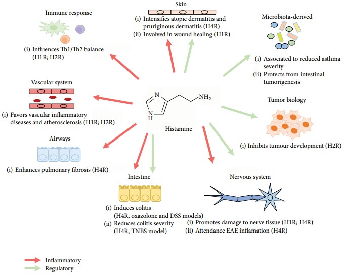 Figure 3. Inflammatory and regulatory functions of histamine on different body sites
(Source: Branco ACCC, et al. 2018)
References
| Target | Cat. No. | Product Name | Size | Species Reactivity | Application | Detection Sample | |
| Histamine | DEIASL281 | Human Histamine ELISA Kit | 96T | Quantitative | Serum, plasma, tissue homogenates and other biological fluids | Inquiry | |
| DEIA2009 | Human his(Histamine) ELISA Kit | 96T | Human | Quantitative | Serum, plasma, tissue homogenates, other biological fluids | Inquiry | |
| DEIA2076 | Histamine Research ELISA Kit | 96T | Quantitative | Animal species, biological fluids | Inquiry | ||
| DEIA1884 | Histamine ELISA Kit | 96T | Quantitative | Plasma, urine | Inquiry | ||
| DEIA052J | Histamine ELISA Kit | 96T | Quantitative | Stool | Inquiry | ||
| DEIABL288 | Histamine Release ELISA Kit | 1 Kit | Quantitative | Heparinized whole blood | Inquiry | ||
| DEIA1962 | Histamine ELISA Kit | 96T | Quantitative | Food | Inquiry | ||
| 1-Methylhistamine | DEIA-XY61 | 1-Methylhistamine ELISA kit | 96T | Quantitative | Urine | Inquiry |
| Target | Cat. No. | Product Name | Expression System | Tag/Conjugate | Application | |
| Histamine | DAG3327 | Histamine [G-BSA] | N/A | G-BSA | IHC, ICC | Inquiry |
| DAG3328 | Histamine [HD-BSA] | N/A | HD-BSA | IHC, ICC | Inquiry | |
| DAGS020 | Histamine Allergen reference material | N/A | N/A | ELISA | Inquiry | |
| DAG-WT1070O | Histamine [BSA] | N/A | BSA | ELISA, LFIA | Inquiry | |
| DAG-WT1070K | Histamine [KLH] | N/A | KLH | Immunogen | Inquiry |
| Target | Cat. No. | Product Name | Host | Isotype | Application | |
| Histamine | DPAb2300 | Anti-Histamine polyclonal antibody | Rabbit | IgG | IHC, IF, IHC-P | Inquiry |
| Histamine | DPAB1723 | Rabbit Anti-Histamine polyclonal antibody | Rabbit | IgG | Dot, IHC, ICC/IF | Inquiry |
| Histamine | DPATB-H82325 | Anti-Histamine polyclonal antibody | Rabbit | IgG | ELISA, IHC-Fr | Inquiry |
| Histamine | DPATB-H83033 | Anti-Histamine polyclonal antibody | Rabbit | IgG | ELISA | Inquiry |
| Histamine | DPAB-DC4352 | Anti-Histamine polyclonal antibody | Rat | IHC, ELISA | Inquiry | |
| Histamine | DMAB8639 | Anti-Histamine monoclonal antibody, clone D98H100 | Mouse | IgA | ELISA | Inquiry |
Loading ......
