
Inquiry Basket
 Loading ......
Globoid cell leukodystrophy or Krabbe disease (KD) (OMIM 245200) is an inherited demyelinating disorder caused by a deficiency of galactosylceramidase (GALC), which leads to the accumulation of its substrates galactosylceramide (GalCer) and galactosylsphingosine (also known as psychosine) in myelin-producing cells. Given that GalCer is readily converted into psychosine via enzymatic diacylation by acid ceramidase (ASAH1) and GalCer is abundantly produced during the active myelination phase of early brain development, the loss of GALC activity is thought to lead to toxic accumulation of psychosine, as well as dysfunction and death of oligodendrocytes and Schwann cells. This results in demyelination in both central and peripheral nervous systems in KD. Although several classifications have been proposed for KD, it can be divided into two subtypes based on the age at symptom onset. Infantile KD (before 12 months) is the most common and fatal form of the disease, which clinically presents with irritability, feeding difficulties, loss of vision and hearing, and seizures after a few months of normal development. Later-onset KD (after 12 months) usually has a better prognosis, and its clinical presentations vary. Genetically, KD is caused by homozygous or compound heterozygous mutations in the human GALC gene, which lead to a dramatic reduction or complete deficit of GALC protein function. In general, symptomatic KD patients have ≤10% of normal GALC activity as measured in dried blood spots, leukocytes or cultured skin fibroblasts. While GALC activity provides a critical index for KD diagnosis, it has also been shown to produce a relatively high false-positive rate in newborn screening. Understanding the relationship between KD genotype and GALC defciency, psychosine accumulation, demyelination, globoid cell pathology and neuroinflammation is an important step in understanding pathogenesis of KD and eventual treatment for KD.
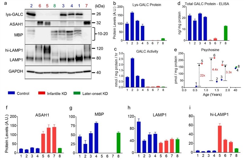 Fig. 1. Biochemical abnormalities in the periventricular white matter of KD.
(Neurobiology of Disease, 2022.)
To examine how the GALC genotypes translate into GALC protein phenotypes, researcher performed activity assay and Western blotting to quantify the function and protein levels of mature, lysosomal GALC (LysGALC) in the PVWM, a major pathological site in KD. Polymorphic variants impair GALC activity by causing reduction in LysGALC protein levels. The loss of GALC activity in KD was associated with absence of Lys-GALC protein in PVWM. Our results show that GALC protein quantification in patient samples can provide information of GALC mutation status.
Psychosine is an important biomarker for diagnosis and newborn screening in KD. To determine how brain psychosine levels are related to disease severity, researcher compared psychosine levels in PVWM of agematched controls and compared them to KD. Our data show a trend of positive correlation between psychosine levels and disease severity. Brain psychosine levels in normal controls increases with age, emphasizing the necessity of using age-matched controls in studies of psychosine in KD.
GALC is a lysosomal enzyme essential for sphingolipid metabolism in myelin-producing cells, including oligodendrocytes and Schwann cells. GALC functional deficiency has been thought to lead to progressive demyelination in the central and peripheral nervous systems. Haploin sufficiency of GALC also causes impaired remyelination. Given the important role of GALC in myelin biology and KD pathogenesis, researcher performed IHC to detect GALC protein in normal human brain tissue. Upon quantification, GALC protein levels were highest levels in PVWM and CC, followed by BA24 white matter, and lowest in parahippocampal white matter in the normal infant brain. The normal adult brain had much higher GALC levels in all white matter regions examined.
Neuroinflammation is thought to play a role in the pathogenesis of KD. To understand the role of neuroinflammation in KD, analyzed the extent and distribution of neuroinflammatory markers, including CD68- positive globoid cells and glial fibrillary acidic protein (GFAP)-positive astrocytes, in white matter of infantile KD. In addition to widespread globoid cells, astrogliosis was prominent in the white matter of KD. GFAP is a specific marker for astrocytes. Although GFAP also labels non-reactive astrocytes, activated reactive astrocytes had elevated GFAP levels that are positively correlated with the extent of astrogliosis. Researcher assessed the burden and distribution of CD4+ and CD8+ T lymphocytes in white matter of KD. Overall, our findings describe and quantify well known and new neuroinflammatory features in the end-stage infantile KD. A severity-dependent increase in globoid cells, astrogliosis and CD8+T cells in various regions of white matter.
 Fig 2. CD8+ T lymphocytes, but not CD4+ T lymphocytes, are present in white matter of infantile KD.
(Neurobiology of Disease, 2022.)
Researcher performed comprehensive biochemical and histopathologic analyses of postmortem brain tissues from KD with different degrees of pathologic severity. Confirmed a gene dose-dependent effect of the p.Ile562Thr variant in brain tissue. GALC protein is highly enriched in oligodendrocytes, which are greatly decreased in end-stage KD brains. Heterogeneity of surviving cell populations in autopsy samples may increase variability in GALC activity. Analysis of non-KD control brain tissues showed age-dependent increases in psychosine in PVWM. In addition to psychosine accumulation, biochemical abnormalities, including increased ASAH1 and hyperglycosylated LAMP1 protein levels, were detected in infantile KD. These increases are likely related to neuroinflammatory response since oligodendrocytes were virtually absent in end-stage KD. ASAH1 levels were increased in infantile KD and were mostly expressed in immune cells. Astrogliosis paralleled globoid cells accumulation and microgliosis, which were found prior to demyelination. These results strongly suggest that differences in neuropathology are related to disease duration. A limitation of our study is the small number of postmortem brain tissues for the analyses, although researcher obtained all available cases in public brain banks. Further statistical analyses need to be done in larger cohorts.
| Target | Cat. No. | Product Name | Host | Isotype | Application | |
| GALC | DPABH-00796 | Anti-GALC (aa 341-591) polyclonal antibody | Rabbit | IgG | WB | Inquiry |
| DCABH-11661 | Anti-GALC monoclonal antibody | Rabbit | IgG | WB, ELISA | Inquiry | |
| CABT-B10301 | Mouse anti-Human GALC monoclonal antibody, clone 3E2 | Mouse | IgG2a | WB, ELISA | Inquiry |
| Target | Cat. No. | Product Name | Size | Application | Detection Sample | |
| GALC | DEIA-FN534 | Human GALC (Galactocerebrosidase) ELISA Kit | 96T | Quantitative | serum, plasma, cell culture supernatants, tissue homogenate | Inquiry |
Loading ......
