The isolation and purification of the target cells of interest have been important research tools in cell biology. Whether it is in vitro stimulation to study cytokines expression profiles or cell co-culture to detect cell function, purity of cells is highly required. Fluorescence-activated cell sorting (FACS) is one of the cutting-edge techniques for isolating and purifying cells, which allows researchers to sort cells according to their physical and chemical properties. FACS enables researchers to isolate single cells with unprecedented 99.99% accuracy, making it the ideal tool for applications where cell purity is critical.
How Does Fluorescence-Activated Cell Sorting Work?
FACS is a type of flow cytometry that uses fluorescent dyes or antibodies to label cells and a flow cytometer to measure their fluorescence and other physical properties. The basic principle of FACS involves passing a cell suspension through a flow cytometer, which uses lasers to excite fluorescent dyes or antibodies and detect emission. By analyzing the fluorescence and other physical properties of each cell, the flow cytometer can sort the cells into different p opulations based on their properties.
FACS is a powerful technique because it allows researchers to analyze and sort cells based on multiple parameters, such as size, shape, intracellular signaling molecules, and surface markers (CD45, CD14, HLA-DR, etc.). This technique has many applications in basic and clinical research, including cell cycle analysis, DNA content analysis, protein expression analysis, cell signaling studies, and cell sorting for further analysis or culture. FACS can also be used to isolate rare or specific cell populations, such as stem cells, tumor cells, or immune cells. This makes FACS a valuable tool for many biological and medical applications, including cell biology, immunology, cancer research, and drug discovery.
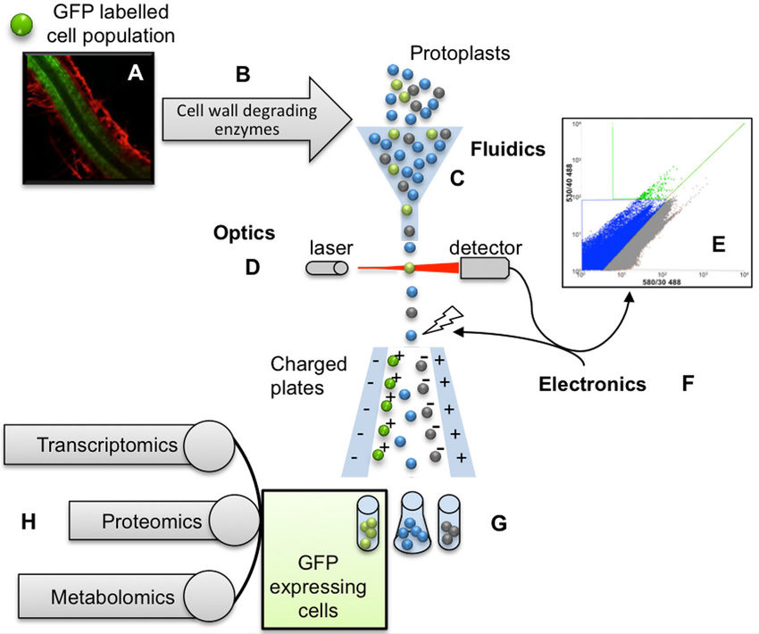 Fig. 1 Fluorescence-Activated Cell Sorting (FACS) workflow. (Carter A D,et al., 2013)
Protocol for Fluorescence Activated Cell Sorting of Live Cells
Materials and Reagents
- Live cells (suspension or adherent)
- Cell culture medium
- Phosphate-buffered saline (PBS)
- Filtered buffer (e.g., FACS buffer, HBSS)
- Fluorescent dyes or antibodies (conjugated to FITC, PE, APC, or other fluorophores)
- Isotype controls (for background subtraction)
- Flow cytometer
- Sterile tubes or plates for cell collection
- Hemocytometer or automated cell counter
| Sample Preparation | - Label cells with fluorescent dyes attached to antibodies that bind to specific cell surface markers. Cells are incubated with fluorescent dyes for 30-60 minutes.
- Wash and dilute cells with buffer solution. The cell sorting buffer contains salts, proteins, and other molecules, which help maintain cell health during sorting. Resuspension cells in PBS to remove unbound dyes.
|
| Instrument Setup | - Calibrate the flow cytometer using calibration beads or cells. Follow the manufacturer's instructions for calibration and optimization of instrument settings, such as laser power, PMT voltage, and compensation.
- Create an experiment in the flow cytometer software program. Choose the appropriate fluorophores and controls for your experiment and set up a gating strategy.
- Run a test sample of unstained or single-stained cells to check instrument performance and adjust the gate and compensation as needed.
|
| Data Acquisition | - Load the cell sample into the flow cytometer and acquire data. Use the appropriate flow rate and sample volume to ensure accurate and reproducible data.
- Flow cytometry uses laser-excited fluorescent dyes and detectors to measure dye fluorescence intensity and classify cells into groups based on their fluorescence intensity and size. The most common sorting methods are forward scattering (FSC) and side scattering (SSC).
- Analyze the acquired data using the flow cytometer software or other data analysis software.
- Calculate cell viability, cell yield, and other parameters of interest based on the gated populations. Compare the results with the appropriate controls and replicate experiments.
|
| Post-Sorting Analysis | - Collect the sorted cells in sterile tubes or plates containing the appropriate cell culture medium or buffer for further analysis.
- Check the sorted cells for purity and viability using a microscope or flow cytometer. Use appropriate staining and controls to confirm the identity and quality of the sorted cells.
- Culture or analyze the sorted cells according to your experimental design. For example, perform functional assays, gene expression analysis, or proteomic analysis on the sorted cells.
|
Note: To maintain cell viability and prevent contamination during subsequent culture, the following measures should be taken during FACS sorting:
- Include serum in buffers.
- Avoid the use of sodium azide, which can be toxic to cells and compromise their viability.
- The experiment should be conducted under aseptic sterile conditions to prevent contamination.
- It is typically not possible to perform intracellular staining before sorting live cells as the permeabilization process required to access intracellular targets would damage the cell membrane and compromise cell viability.
Performing FACS requires careful preparation, optimization, and analysis of experimental procedures. By following the tips and guidelines provided in this article, researchers can optimize their FACS experiments and obtain reliable and reproducible results. As a leading provider of innovative tools and services for life science research, Creative Diagnostics offers a wide range of products for FACS analysis, including fluorescent dyes and antibodies.
Reference
- Carter A D, Bonyadi R, Gifford ML. The use of fluorescence-activated cell sorting in studying plant development and environmental responses. The International journal of developmental biology, 2013, 57: 545-552.