Epigenetics refers to heritable but reversible changes in gene expression that occur without altering the underlying DNA sequence. The major mechanisms include DNA methylation, histone modifications, chromatin remodeling, and non-coding RNAs. Together, these modifications orchestrate when and how genes are turned on or off. Importantly, epigenetics explains how environmental influences such as diet, stress, and lifestyle can shape gene activity, providing critical insights for disease diagnosis, prevention, and therapy. To explore these mechanisms, researchers have developed a broad toolkit of methods that allow precise mapping of epigenomic landscapes and dynamic regulation.
Fig. 1 Overview of epigenetic regulatory mechanisms
DNA-Level Methods: Mapping Methylation
DNA methylation is one of the most extensively studied epigenetic marks, typically associated with gene silencing. Several technologies enable genome-wide profiling of methylation states.
Bisulfite sequencing (WGBS, RRBS) is a well-established method for detecting methylated cytosines in genomic DNA. The key idea of this method is combining the power of high-throughput DNA sequencing with the treatment of DNA with sodium bisulfite. When exposed to sodium bisulfite, unmethylated cytosines are converted to uracils, whereas methylated forms of cytosine remain unchanged. The location of the methylated cytosines is determined by comparing treated and untreated sequences. This method is widely applied in identifying biomarkers for cancer, imprinting disorders, and developmental abnormalities.
Fig. 2 Conversion by bisulfite treatment
Methylation arrays enable quantitative interrogation of CpG sites methylation across the genome, offering high-throughput capabilities that minimize the cost per sample. They are frequently used in population-level research to detect disease-associated epigenetic changes.
MeDIP-seq and MBD-seq are affinity-based enrichment methods. MeDIP-seq (methylated DNA immunoprecipitation sequencing) employs a 5-methylcytosine monoclonal antibody to recover methylated fragments from single-stranded DNA, whilst MBD-seq captures double-stranded methylated DNA fragments using the methyl-binding domain from MECP2. Both results are generally concordant but non-identical. Both approaches risk poor coverage of the medium to low CpG density regions of the genome, and neither allows for single-base CpG resolution. Both detect 5-methylcytosine exclusively, unlike bisulfite conversion, which does not differentiate between 5-methylcytosine and 5-hydroxymethylcytosine. MeDIP-seq and MBD-seq allow researchers to profile methylated regions across multiple samples with lower cost, supporting comparative analyses in oncology and toxicology studies.
Fig. 3 Comparison of MeDIP-seq and MBD-seq methods
Aberrant methylation has been associated with the majority of diseases, including cancer, neurodegenerative, cardiovascular and autoimmune disorders. Through these tools, DNA methylation analysis has become a cornerstone for precision diagnostics, early cancer detection, and monitoring epigenetic therapies.
Histone proteins carry diverse chemical modifications - including acetylation, methylation, phosphorylation, and ubiquitination - that collectively shape chromatin accessibility and gene regulation.
ChIP-seq (Chromatin Immunoprecipitation sequencing) combines ChIP with next-generation sequencing. This method uses an antibody for a specific DNA-binding protein or a histone modification to identify enriched loci within a genome. It has been instrumental in defining active promoters, enhancers, and repressive domains.
CUT&RUN and CUT&Tag are two novel technologies. They are performed in situ on immobilized, intact cells without crosslinking. DNA fragmentation is achieved using either micrococcal nuclease (MNase) for CUT&RUN or a hyperactive transposase (Tn5) for CUT&Tag fused to Protein A and/or Protein G. The fusion protein is directed through binding of the Protein A/G moiety to the Fc region of an antibody bound to the target of interest. The DNA under the target is subsequently cleaved and released and the fusion protein-antibody-chromatin complex is free to diffuse out of the cell. DNA cleavage products are extracted and then processed by next-generation sequencing (NGS). CUT&RUN and CUT&Tag represent next-generation approaches that require fewer cells and yield higher resolution, enabling applications in clinical samples and even single-cell studies.
Fig. 4 Comparison of ChIP, CUT&RUN, and CUT&Tag assays
Mass spectrometry (MS) allows determining with high accuracy the mass (or more precisely the mass/charge ratio, m/z) of different types of molecules, including peptides and proteins. PTMs are identified by MS and localized to specific residues by detecting a "delta-mass" between the theoretical and experimentally measured masses of peptide/proteins. As a consequence, theoretically any PTM or combination of PTMs can be profiled in a single run, without requiring a priori knowledge of the type or site of the modification, and in a highly quantitative manner. MS-based histone proteomics provides a global view of histone post-translational modifications, supporting drug discovery efforts by revealing combinatorial histone codes.
These methods empower researchers to link specific histone modifications with gene expression programs, uncovering regulatory networks in cancer, immune disorders, and neurodegenerative diseases. They are also essential tools in epigenetic drug development, where inhibitors of histone-modifying enzymes are actively being explored as therapies.
Chromatin Accessibility and 3D Genome Architecture
Beyond DNA and histones, the three-dimensional structure of the genome plays a critical role in regulation. Technologies that probe chromatin accessibility and spatial genome organization are key to understanding enhancer–promoter interactions and higher-order chromatin folding.
DNase-seq and FAIRE-seq are techniques used to identify regulatory sites across the genome. DNase-seq employs the DNase I enzyme to preferentially digest nucleosome-depleted regions, known as DNase I hypersensitive (HS) sites. On the other hand, FAIRE-seq enriches nucleosome-depleted DNA by using formaldehyde fixation followed by phenol-chloroform extraction. However, these earlier methods have largely been replaced by ATAC-seq in modern workflows.
ATAC-seq (Assay for Transposase-Accessible Chromatin) is a popular method for determining chromatin accessibility across the genome. ATAC-seq uses hyperactive Tn5 transposase to simultaneously cut and ligate adapters for high-throughput sequencing at regions of increased accessibility. It widely used in mapping regulatory elements and defining cell-type–specific signatures.
Hi-C and related 3C-based methods reveal the three-dimensional organization of chromatin, including topologically associated domains (TADs) and long-range enhancer–promoter loops. Chromosome conformation capture (3C) is the preferred method to study chromatin interactions. 3C determines chromatin conformation by analyzing interaction frequencies between selected genomic sites. Many alterations of the 3C principle have been developed for large-scale applications including circular chromosome conformation capture (4C), chromosome conformation capture carbon copy (5C), chromatin interaction analysis by paired-end tag sequencing (ChIA-PET) and Hi-C. All of these methods generated complementary evidence that the formation of chromatin loops allows interaction between genes and regulatory elements and led to great advances in our understanding of chromatin and genome organization. These insights are critical for understanding structural variants in cancer genomes and developmental gene regulation.
Fig. 5 Principles of 3C-derived methods
By integrating accessibility and 3D genome data, scientists can link distal regulatory elements to their target genes, offering powerful insights into complex diseases and regulatory mutations.
Dynamic and Interaction-Based Methods
Epigenetics is not static - it is dynamic and responsive to external stimuli. The epigenetic processes that modulate access to DNA in response to upstream signals include DNA methylation, covalent modification of histones, nucleosome remodeling, nuclear dynamics and chromatin interaction with regulatory noncoding RNAs. Techniques that capture these dynamics provide unique insights.
Live-cell imaging allows for the observation of chromatin movement and epigenetic regulator activity in real time within living cells. Using fluorescently labeled histones, transcription factors, or epigenetic enzymes, this technique offers direct visualization of gene activation, chromatin condensation, or remodeling events under natural conditions. It is particularly useful for studying how drugs or environmental factors influence chromatin states over time.
Proximity labeling approaches (e.g., BioID, APEX, TurboID) enable mapping of protein-protein interactions within chromatin complexes. By fusing an engineered enzyme to an epigenetic regulator, nearby proteins are biotinylated and subsequently identified by mass spectrometry. This approach is particularly valuable for uncovering the composition of chromatin remodeling complexes or histone modifier interactomes in their native environment. Unlike classical co-immunoprecipitation, proximity labeling captures weak or transient interactions that may otherwise be missed. These methods provide a high-resolution view of epigenetic networks and how they rewire under stress, disease, or drug treatment.
Fig. 6 Comparison of BioID and APEX proximity labeling methods
CRISPR/dCas9-based epigenome editing allows targeted manipulation of DNA methylation or histone modifications without altering the underlying DNA sequence. By fusing dCas9 to effectors such as TET demethylases or histone acetyltransferases, researchers can precisely activate or repress specific genes. This approach is widely applied to functionally validate candidate regulatory elements identified by sequencing methods, as well as to model disease-associated epigenetic changes. Beyond research, dCas9-based epigenome editors hold therapeutic potential, offering a way to reprogram abnormal gene expression in cancer, neurological disorders, or metabolic diseases.
Epigenetic modifications play an important role in regulating gene expression. Understanding different epigenetic states is crucial for dissecting the gene regulatory mechanisms. These approaches bridge the gap between static maps and dynamic processes, helping researchers model disease mechanisms and test epigenetic interventions.
Conclusion
Epigenetics provides a framework for understanding how gene activity is regulated beyond DNA sequence alone. With rapid development in the epigenetics field, many powerful techniques have been discovered to access epigenetic functions and mechanisms of the epigenome and its associated proteins. Each technique offers unique advantages, collectively enabling breakthroughs in disease diagnosis, therapeutic discovery, and precision medicine.
References
Smigielski L, et al. Epigenetic mechanisms in schizophrenia and other psychotic disorders: a systematic review of empirical human findings[J]. Molecular psychiatry, 2020, 25(8): 1718-1748.
Neary J L, et al. Comparative analysis of MBD-seq and MeDIP-seq and estimation of gene expression changes in a rodent model of schizophrenia[J]. Genomics, 2017, 109(3-4): 204-213.
Li Y. Modern epigenetics methods in biological research[J]. Methods, 2021, 187: 104-113.
Su X, et al. Mass spectrometry-based strategies for characterization of histones and their post-translational modifications[J]. Expert review of proteomics, 2007, 4(2): 211-225.
Wu Y L, et al. Epigenetic regulation in metabolic diseases: mechanisms and advances in clinical study[J]. Signal Transduction and Targeted Therapy, 2023, 8(1): 98.

 Loading ......
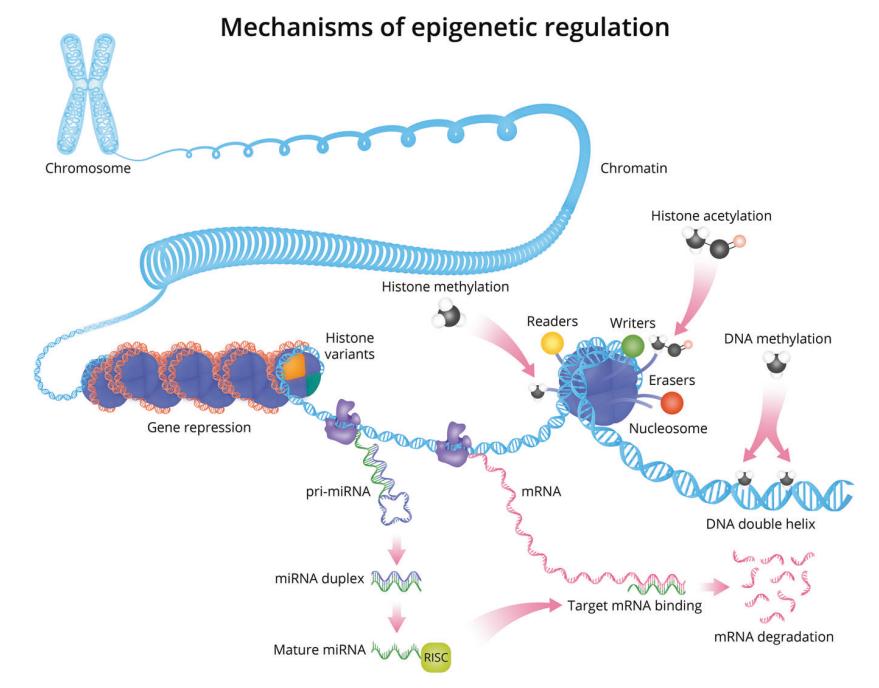 Fig. 1 Overview of epigenetic regulatory mechanisms
 Fig. 2 Conversion by bisulfite treatment
 Fig. 3 Comparison of MeDIP-seq and MBD-seq methods
 Fig. 4 Comparison of ChIP, CUT&RUN, and CUT&Tag assays
 Fig. 5 Principles of 3C-derived methods
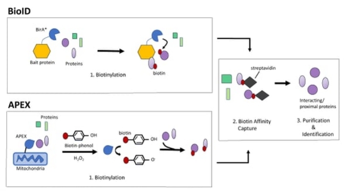 Fig. 6 Comparison of BioID and APEX proximity labeling methods