
Inquiry Basket
 Loading ......
Enterovirus (EV) mainly transmitted through the fecal-oral route and targets the gastrointestinal epithelium at the early stage of its life cycle. In addition, it may be transmitted through respiratory tract, and some EVs such as EV D68 are transmitted through this route preferentially. It is a genus included fifteen species, enterovirus A-L and rhinovirus A-C. People of all ages can be infected with EVs, infection ranges from acute and self-limited febrile diseases to meningitis, endocarditis, hepatitis and acute flaccid myelitis. Poliomyelitis is the most serious disease caused by enterovirus (poliovirus, PV).
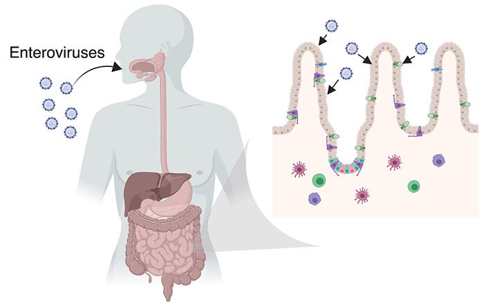 Fig. 1 Transmission route of Enterovirus (Wells AI, et al. 2019)
Poliovirus, Coxsackievirus and Echovirus are all belonging to Enterovirus genus. Coxsackievirus was merged into EV A, B and C strains. According to its pathogenicity in newborn mice, it is divided into group A and group B. Group A Coxsackievirus tends to infect skin and mucosa, while group B Coxsackie virus tends to infect heart, pleura, pancreas and liver. Echoviruses include 34 strains, most of which belong to Enterovirus A, B and C except echovirus 10, 22 and 23.
Enterovirus is a genus of non-enveloped positive single-stranded RNA virus, belonging to Picornaviridae family. The virus particles are icosahedral particles with a diameter of approximately 30 nm. The protein capsid consists of VP1, VP2, VP3 and VP4 proteins. VP1, VP2 and VP3 appear in the outer layer of the capsid, and VP4 is the internal protein. There is a 7.5kb virus genome in the capsid. Genome is a linear molecule of single-strand RNA, containing an open reading frame (ORF) with 3 '(~100 bases) and 5' (~800 bases) terminal untranslated regions (UTR) on both sides. ORF contains genes encoding 11 proteins. There are four capsid proteins (VP 1-4), two viral proteases (2A and 3C), an RNA-dependent-RNA-polymerase (3D), two proteins involved in RNA synthesis (2B and 2C), RNA synthesis primer (3AB) and a small peptide (VPg) of 20-24 amino acids derived from gene 3B.
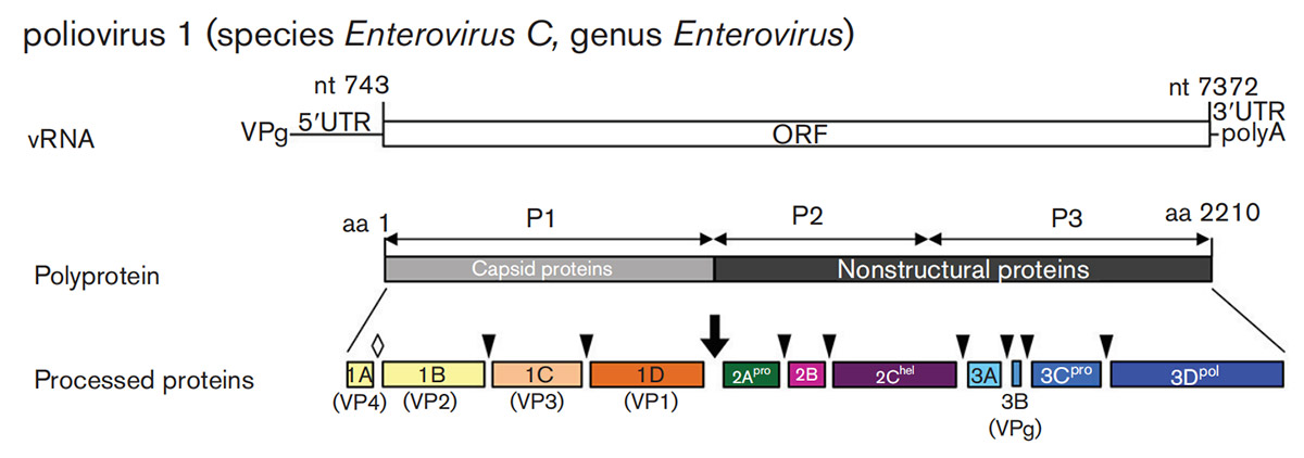 Fig. 2. Genome organization and expression of enteroviruses (Zell R, et al. 2017)
Enterovirus enters host cells by binding to cell surface receptors and undergoing receptor-mediated endocytosis. Then enteroviruses are shelled to release the viral genome. The viral RNA enters the cytoplasm and is translated by the host ribosome. The newly synthesized genome is packaged into virus particles by capsid proteins VP0, VP1 and VP3. Finally, new progeny viruses are released in the vesicles in non-lytic form or during cell lysis.
 Fig. 3 Enterovirus life cycle (Wells AI, et al. 2019)
For patients, timely laboratory diagnosis of EV infection may reduce the use of antibiotics, shorten hospital stay, and minimize the risk of complications. For researchers, laboratory diagnosis of EV infection can also help evaluate the effectiveness of potential therapies such as new antiviral or immunotherapy, and provide clues for further development of vaccines.
Non-polio enteroviruses usually cause mild infections and require only over-the-counter cold medicines. Three vaccines against EV71 have been licensed in China. The effectiveness of the three vaccines after two doses of immunization ranged from 90.0% to 97.4% after one year of monitoring to 95.1% after two years of follow-up. Other vaccines, such as recombinant and other forms of subunit vaccines, vector vaccines and viroid particle vaccines, are in the early stage of development. In addition, consideration is being given to developing vaccines targeting Coxsackie A16, A6 and A10 to develop combination/multivalent vaccines.
Loading ......
