
Inquiry Basket
 Loading ......
Prostate Cancer (PCa) is one of the leading causes of cancer-related deaths among men, and the major cause of death from PCa is metastasis. Many factors have been implicated in this process, but the precise mechanisms for the directional migration and invasion of tumor cells into specific organs is unknown. Chemokines and their corresponding receptors appear to play an important role in the directional migration of hematopoietic cells to specific anatomical sites. Recent studies have shown similar to their role leukocyte trafficking, chemokines and chemokine receptors are involved in cancer metastasis. The chemokine receptor CXCR5 is primary expressed by the B cells and certain T cells and controls their migration into and within lymph nodes. However, recent studies have shown that CXCR5 is expressed by the colon carcinoma and mediates metastasis to the liver. Others have shown relatively higher CXCR5 mRNA expression by some PCa cell lines. At the same time, CXCR5 antibody plays an important role in characterizing the functional significance of CXCR5.
Prostate TMAs consisting of tissue from PCa, BPH and NM cases were evaluated for CXCR5 expression. Prostate tissues from PCa, BPH and NM patients were stained with isotype control (negative stain control) or anti-CXCR5 antibodies. The results showed that the expression of CXCR5 in PCa cases was significantly higher than that in NM tissues (p < 0.001). PCa tissues displayed membrane, cytoplasmic as well as nuclear localization of CXCR5. PCa tissues with Gleason scores ≥ 7 displayed predominantly nuclear and to a lesser degree membrane and cytoplasmic CXCR5 expression patterns. PCa cases with Gleason scores ≤ 6 for the most part displayed membrane and cytoplasmic, but minimal nuclear, CXCR5 expression patterns. BPH and NM tissues had overall lower reactivity and displayed membrane expression patterns.
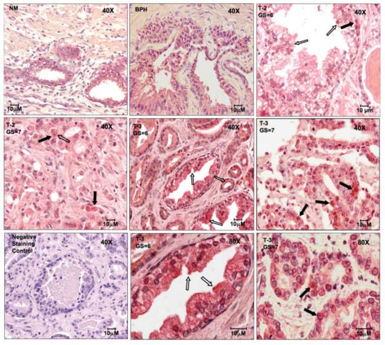 Fig1. CXCR5 expression by prostatic tissues.
(Source: Int J Cancer, 2009)
The functional significance of CXCR5 expression by PCa cells was demonstrated by the ability of LNCaP and PC3 cell lines to migrate toward CXCL13. LNCaP and PC3 cell lines were co-cultured with 1.0 μg/ml of anti-CXCR5 antibody or isotype control monoclonal antibody during migration assays using CXCL13. Similarly, LNCaP and PC3 cells were co-cultured with 1.0 μg/ml of anti-CXCR5 antibody or isotype control monoclonal antibody during invasion across Matrigel in response to CXCL13. The results showed that both PC3 and LNCaP cell lines migrated to CXCL13, but LNCaP cells displayed a higher migration potential than PC3 cells. The addition of mouse anti-human CXCR5 antibody significantly neutralized this chemokine receptor-dependent motility. This CXCL13-mediated invasion was significantly reduced by CXCR5 blockade.
PC3 and LNCaP cell lines were able to invade Matrigel by degrading it’s constituents. LNCaP, PC3 and PrEC cells were cultured for 16 hours with and without CXCL13 (100 ng/ml) or anti-CXCR5 antibody (1 μg/ml). Total RNA was isolated, and quantitative RT-PCR analysis was performed in triplicate to quantify collagenase and gelatinase mRNA expression; copies of mRNA transcripts are expressed relative to actual copies of 18S rRNA (upper panel). The results suggest that PC3 cells are predisposed to collagenase(s) production without CXCL13 induction, but can be induced to significantly increase their production of MMP-1, but not MMP-13, after CXCL13 treatment. The findings indicated that CXCL13-CXCR5 interactions modestly increased gelatinase mRNA expression by PCa cells, but significantly enhanced active MMP-2 and -9 production by these malignant cells. Taken together, these data show that CXCL13 modulates stromelysin expression (and activation) in a CXCR5-dependent fashion.
Mounting evidences suggests that multiple chemokines and their corresponding receptors are involved in PCa progression and play significant roles in metastatic process. The use of CXCR5 antibody plays a crucial role in this process. In conclusion, the evidences show that PCa cells and tissues significantly express CXCR5. While increased CXCR5 expression by PCa cases suggests it plays a role in invasion, it is also plausible that the reduction in CXCR5 surface expression in advanced cases promotes less adhesion. This could potentially allow tumor cells to escape primary sites and spread. To explain, CXCR5 activation leads to integrin co-aggregation to induce adhesion to (bone marrow) endothelial cells, since integrins co-aggregate with CXCR5 and other chemokine receptors following activation to promote firm cell adhesion. Indeed, CXCL13 is an arrest chemokine for B cells in high endothelial venules. Alternatively, the observation of CXCR5 expression transitioning from the surface to nuclei during PCa progression might also be due to receptor internalization and/or nuclear translocation following CXCL13 stimulation.
| Target | Cat. No. | Product Name | Host | Isotype | Application | |
| CXCR5 | DMABT-H29518 | Magic™ Anti-CXCR5 monoclonal antibody, clone SG9C3 [Alexa Fluor® 488] | Rat | IgG2b | FC | Inquiry |
| DMABT-H25041 | Anti-CXCR5 monoclonal antibody, clone NV6VCFF [PE] | Mouse | IgG2b | FC | Inquiry | |
| DMABT-H22012 | Mouse anti-CXCR5 Monoclonal antibody [clone 98.2], PE-Conjugated | Mouse | IgG1 | Inquiry | ||
| DMABT-H16745 | Anti-CXCR5 monoclonal antibody, clone 62606 [FITC] | Mouse | IgG2b | FC | Inquiry | |
| CABT-BL1172 | Anti-CXCR5 monoclonal antibody, clone 62606 | Mouse | IgG2b | BL, Neut, FC, IHC, ICC | Inquiry | |
| CABT-BL7446 | Anti-Cxcr5 Monoclonal antibody, clone TQSDM6 [Biotin] | Rat | IgG2a, κ | FC | Inquiry | |
| CABT-BL7447 | Anti-Cxcr5 Monoclonal antibody, clone TQSDM6 [PerCP-eFluor® 710] | Rat | IgG2a, κ | FC | Inquiry | |
| DMABT-H29518U | Rat Anti-Human CXCR5 Monoclonal Antibody, clone SG9C3 | Rat | IgG2b, κ | FC | Inquiry | |
| CABT-L1264 | Rabbit Anti-Human CXCR5 monoclonal antibody, clone 7I46M4 | Rabbit | IgG | WB | Inquiry |
| Target | Cat. No. | Product Name | Species Reactivity | Application | Detection Sample | |
| CXCR5 | DEIA-FN349 | Human CXCR5 (C-X-C chemokine receptor type 5) ELISA Kit | 96T | Quantitative | serum, plasma, cell culture supernatants, tissue homogenate | Inquiry |
Loading ......
