
Inquiry Basket
 Loading ......
Circulating tumor cells (CTCs) are tumor cells that have sloughed off the primary tumor and extravasate into and circulate in the blood. Although CTCs originate from the primary tumor, they are distinct from primary tumor cells, with EMT transition properties that help them break free from the primary tumor and facilitate intravasation into the bloodstream, dissemination in clusters of CTCs to increase metastatic potential, and exhibit stemness features that enhance their ability to initiate metastasis. Understanding of the metastatic cascade of CTCs has tremendous potential for the identification of targets against cancer metastasis. The enumeration of CTCs has emerged as a simple and effective biomarker with many applications in cancer research for prognosis and treatment. Moreover, they represent an entire cancer-derived cell population, providing a potent tool to study tumor heterogeneity and progression of the disease at various stages.
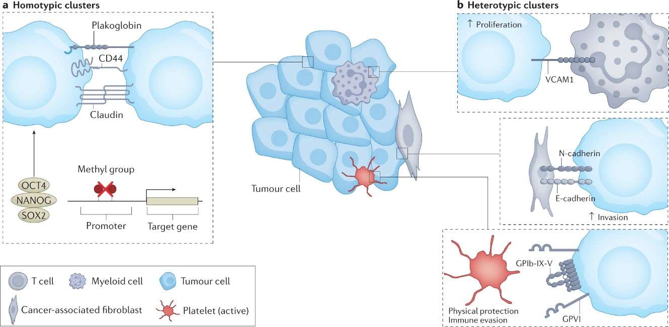 Fig. 1: Biological features of circulating tumor cell clusters
When transported in the bloodstream, a major of CTCs are constrained by detrimental shear stress. Only a small fraction of CTCs interacts tightly with platelets, neutrophils, macrophages, myeloid-derived suppressor cells (MDSCs), or cancer-associated fibroblasts (CAFs) to escape the immune system and promote their survival. Tumor-associated macrophages (TAMs) contribute to the extravasation of CTCs. CTCs was found to be able to induce monocyte differentiation to TAMs, and the feedback loop between TAMs and cancer cells is essential for the EMT program of CTCs and intravasation into the blood stream. Neutrophils can directly adhere to CTCs through the Mac-1/ICAM-1 interaction and act as a bridge between tumor cells and the liver parenchyma, thus promoting extravasation and liver metastasis. CTCs clusters with neutrophils anchor to the vascular endothelium for extravasation while resisting shear stress, and the process is mediated by a series of cell adhesion proteins, such as cadherin, integrin, and surface glycoprotein.
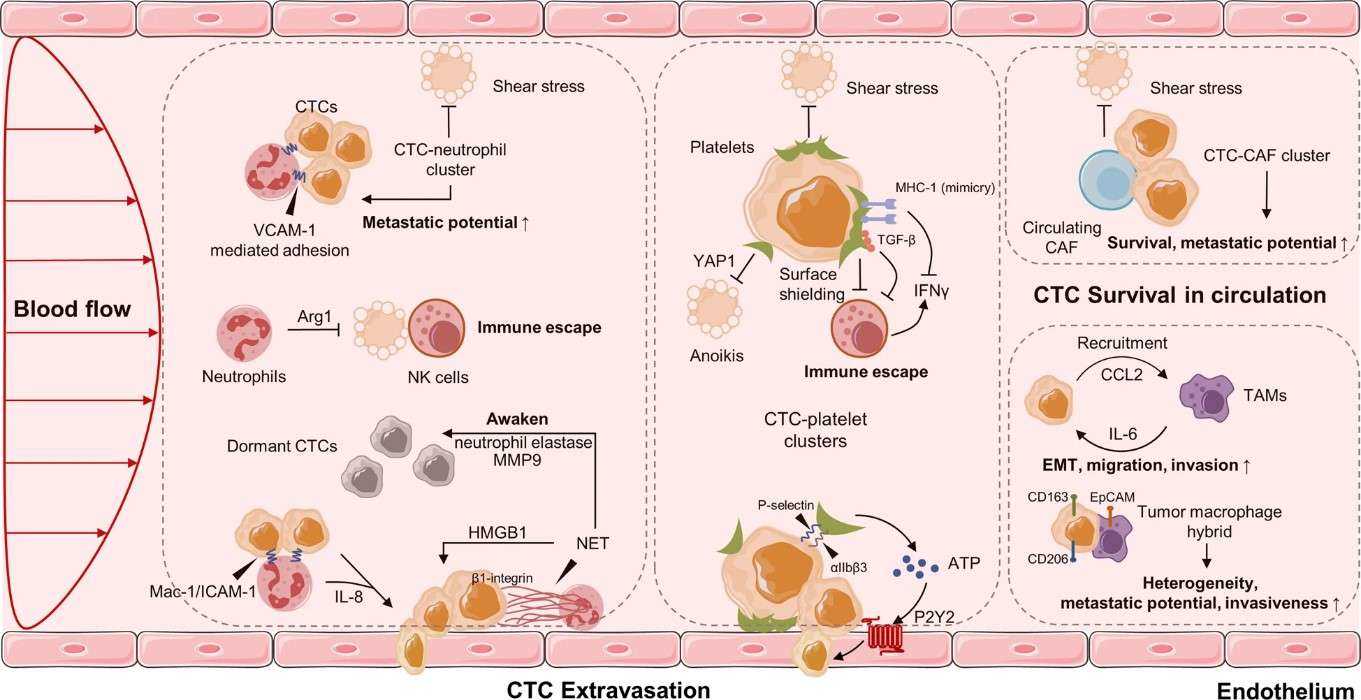 Fig. 2: CTCs in the blood microenvironment, and their interaction with neutrophils, platelets, CAFs and TAMs.
EMT provides a potential mechanistic basis for how CTC intravasate in primary tumors to reach the circulation, and seed tumor implants at distant secondary sites. EMT is associated with the increased tumor cell motility and an invasive phenotype. These changes are typically characterized by loss of E-Cadherin expression and subsequent translocalization of β-catenin from the cell membrane into the nucleus, increased expression of vimentin, production of matrix metalloproteinase enzymes, and upregulation of various EMT-inducing transcription factors such as Twist, Snail, and Slug. In a study of metastatic breast cancer there was at least one of three EMT markers (Akt2, PI3K, and Twist1), assessed by RT-PCR, to be expressed by the CTC population in 62% of patients harboring CTC. Evaluating CTC expression of EMT markers Twist and vimentin by immunofluorescence, it has been found vimentin/Twist expressing CTC in 77% of early-stage breast cancer patients compared to 100% of patients with metastatic disease.
As most CTCs perish in the circulation, and only limited CTCs survive and infiltrate distant organs. They are a very minor fraction of blood cells, as there is usually less than 10 CTCs in 1 ml of blood. Isolating these very rare CTCs from the massive pool of circulating blood cells is a big technical challenge. CTC markers help to identify and isolate these very rare cells.
The most common marker used for CTCs is EpCAM. EpCAM-based CTC detection technologies are widely applied for cancers that strongly express EpCAM, such as breast and prostate cancer, pancreatic, colorectal, and hepatocellular cancers. The presence of these EpCAM-positive CTCs predicts early distant metastasis and poorer survival of patients. In addition, after leaving the primary tumor, circulating tumor cells reshape their integrin expression profile to avoid apoptosis connected with lack of attachment, or to escape immune system surveillance, including decreased expression of epithelial markers (E-cadherin, ZO-1, claudins, and occludins) and increased expression of mesenchymal markers (vimentin, N-cadherin, fibroblast-specific protein1, and fibronectin). Other biomarkers, such as human epidermal growth factor receptor-2 (HER2), estrogen receptor, prostate-specific membrane antigen, folate receptor, and survivin, have been described as CTCs markers in different cancers. Most of these cancer-specific CTC markers are in accordance with the specific molecular markers of the primary tumor. However, it should be mentioned that for melanoma, the detection technologies of CTCs are based on several melanoma cell adhesion molecules, such as HMW-MAA, MART-1, CD146, and MAGE A3, which are very specific molecular markers for melanoma.
As CTCs are very heterogeneous population, for their detection it is better to use pan-reactive antibodies. On the other hand, when trying to identify a particular source of circulating tumor cells, specific combinations of anti-cytokereatin antibodies are needed. Creative Diagnostics has developed a portfolio of CTCs antibodies can be used for efficient capture, recovery, and analysis of CTCs from liquid biopsy samples, cell culture, and other sample types, to facilitated the clinical applications of CTCs in cancer screening, treatment response monitoring, and prognosis evaluation.
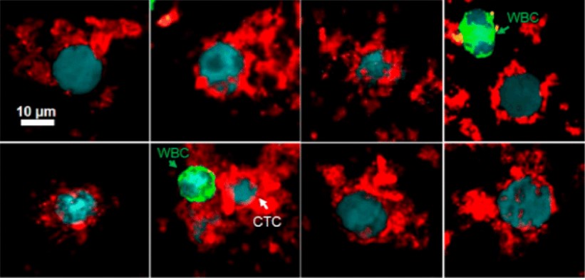 Fig. 3: Fluorescent images of eight CTCs (Hoechst 33342+, F-MNPs+, CD45−) and WBCs (Hoechst 33342+, CD45+) from breast cancer patients' blood samples.
Ref: Hepel, Maria. "Magnetic nanoparticles for nanomedicine." Magnetochemistry 6.1 (2020): 3.
 Fig. 4: Detection of Circulating tumor cells (CTCs) in renal cell carcinoma samples (RCC patient.
Ref: Zhu, Peixuan, et al. "Detection of tumor-associated cells in cryopreserved peripheral blood mononuclear cell samples for retrospective analysis." Journal of translational medicine 14 (2016): 1-12.
| Cancer types | Epithelial markers | Mesenchymal markers | Specific markers |
| Breast cancer | EpCAM | Vimentin | HER2 |
| CK5 | Twist | ER | |
| E-Cadherin | Fibronectin | AR | |
| CK7 | N-Cadherin | MRP | |
| CK8 | SERPINE1/PAI1 | ||
| CK18 | β-catenin | ||
| CK19 |
| Cancer types | Epithelial markers | Mesenchymal markers | Specific markers |
| Non-small-cell lung cancer | CK7 | Vimentin | Folate receptor |
| CK8 | Twist | Telomerase activity | |
| CK18 | N-Cadherin | ||
| CK19 | AXL | ||
| EpCAM |
Loading ......
