
Inquiry Basket
 Loading ......
The tumour microenvironment, also termed the tumour stroma or tumour mesenchyme, includes fibroblasts, immune cells, blood vessels and the extracellular matrix and substantially influences the initiation, growth and dissemination of gastrointestinal cancer. Cancer-associated fibroblasts (CAFs) are one of the critical components of the tumour mesenchyme and not only provide physical support for epithelial cells but also are key functional regulators in cancer, promoting and retarding tumorigenesis in a context-dependent manner.
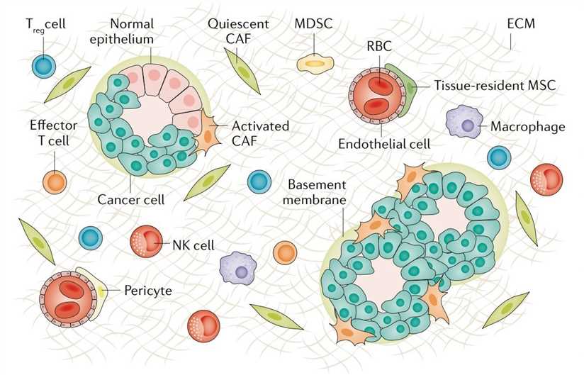 Fig. 1 | Cellular components of the tumour microenvironment.
(Source: Nature reviews. Gastroenterology & hepatology,2019)
Fibroblasts are spindle-shaped, non-epithelial and nonimmune cells embedded in the ECM that are easily propagated in adherent cell culture. They are a major constituent of the stroma in gastrointestinal organs, and as in other tissues, they are highly organized. Throughout the gastrointestinal tract, a reticular network of stromal cells lies coincident with the epithelial basement membrane. The subepithelial plexus, composed of reticular stromal cells, entirely surrounds the glandular axis from the stomach to the rectum. This compartment is dynamic, with a radial axis of proliferation and differentiation, analogous to the epithelium, developing from gremlin 1-expressing intestinal reticular stem cells. Functionally, fibroblasts are fundamental regulators of ECM synthesis and of paracrine and juxtacrine signalling to nearby epithelium to regulate growth and differentiation, and they are also ready to respond to tissue injury, either in wounding or tumorigenesis. CAFs are generally accepted to be all of the fibroblasts found within and surrounding a cancer. This group includes native, normal fibroblasts and activated, proliferating (Ki67+) or recruited fibroblasts in response to stimuli from cancer.
Marker heterogeneity. Representative CAF markers include but are not limited to αSMA, the serine protease fibroblast activation protein (FAP), fibroblast-specific protein 1 (FSP1), platelet-derived growth factor receptor-α (PDGFRα) and PDGFRβ. It is possible that CAFs identified by a single marker are composed of a range of distinct CAF subtypes that have functionally opposing roles in cancer progression.
Functional heterogeneity. Studies suggest that CAFs are composed of various functionally heterogeneous subsets that either promote or restrain cancer growth. CAFs are more than inert cells; they actively modulate their environment.
Intratumoural heterogeneity. Analogous to phenotypic heterogeneity among cancer cells, CAF phenotypes are different not only between tumours (intertumoural heterogeneity) but also within tumours (intratumoural heterogeneity).
Although studies have begun to illustrate the heterogeneous nature of CAFs, little is known about the origins of CAFs. Different pathways probably exist for the development of different CAF subpopulations. Fundamentally, cancer develops within an initially normal organ. Depending on the stage of tumorigenesis, there will, at least in very early stages, exist some remnant native fibroblasts. Thereafter, these cells are increasingly replaced by new CAFs that are different from native fibroblasts within normal tissue.
Stromal and epithelial interactions. CAFs directly confer growth advantages to cancer cells via paracrine signalling, exosome transfer and physical interaction.
Tumour immunology. CAFs are major contributors to an immunosuppressive TME that might act as a restitution programme to help support the epithelium in acute injury but promotes cancer growth in a tumour setting.
Angiogenesis. Neovascularization in cancer is regulated not only by tumour cells but also by stromal cells. Indeed, CAFs promote tumour angiogenesis directly by secreting pro-angiogenic factors and indirectly by producing ECM.
Despite advances in chemotherapy, molecularly targeted drugs and immunotherapy, these treatments offer survival benefits only to a small group of patients. Emerging evidence has demonstrated that CAFs confer substantial resistance to cancer therapeutics via impaired drug delivery and biochemical signalling. The ECM produced by CAFs acts as a physical barrier to prevent the penetration of drugs by increasing interstitial fluid pressures and inducing vascular collapse. In regards to molecularly targeted drugs, HGF and IGF2 released by CAFs contribute to the resistance to tyrosine kinase inhibitors. Furthermore, CAFs influence the responsiveness to immune checkpoint inhibitors by shaping the immunosuppressive TME. The radioresistance is attributed, in part, to an altered interaction between cancer cells and CAFs following radiation.
Clinical implementation Biomarkers In addition to the functional contribution of CAFs to cancer progression described above, CAFs and their gene expression patterns have diagnostic and prognostic value in clinical oncology. Gene expression analyses and proteome profiling of cancer tissues have revealed that stromal gene signatures predict poor patient outcome in multiple types of gastrointestinal cancer. Because CAFs accumulate at the tumour site at an early stage of tumorigenesis, future investigations will probably identify valuable CAF markers that might facilitate early detection of cancer.
CAFs are important in the development of gastrointestinal cancers, both in their promotion and,in their antagonism. CAFs are not one entity but rather contain heterogeneous functional subpopulations including pCAFs, rCAFs and probably also a neutral subset that neither promotes nor retards (nCAFs). CAF biology is mediated through the direct and paracrine interactions of CAFs with both cellular (tumour cells, immune cells and vascular cells) and acellular (ECM) compartments.
Reference
| Target | Cat. No. | Product Name | Host | Isotype | Application | |
| Epithelium | DMABT-49595MH | Anti-Fibroblasts/Epithelial Cells monoclonal antibody, clone E8-GJC [R-PE] | Mouse | IgG2a | FC | Inquiry |
| FAP | CABT-L3217 | Mouse Anti-Mouse Fibroblast Activation Protein monoclonal antibody, clone 84.4 | Mouse | IgG1 | WB, ELISA, IP | Inquiry |
| Fibroblast | DMABT-H21103 | Anti-Fibroblasts monoclonal antibody, clone UF-8 | Mouse | IgG1 | FC, IF, IHC | Inquiry |
| DMABT-Z59721 | Anti-Fibroblast monoclonal antibody, clone 7D7 | Mouse | IgG1 | IHC-Fr, FC, ICC, IF, IHC-P | Inquiry | |
| Fibroblast Surface Protein | DMAB13751 | Anti-Fibroblast Surface monoclonal antibody, clone 2C20 | Mouse | IgM | WB, IHC | Inquiry |
| DMABT-Z59720 | Anti-Fibroblast Surface Protein monoclonal antibody, clone 3D22 | Mouse | IgM | ICC, IF, IHC-Fr, WB | Inquiry | |
| Myofibroblasts | DMABT-Z59834 | Anti-Myofibroblasts monoclonal antibody, clone RU 4F5 | Mouse | IgG1 | IHC-Fr | Inquiry |
| Reticular Fibroblasts and Reticular Fibres | DMABT-Z59940 | Anti-Reticular Fibroblasts and Reticular Fibres monoclonal antibody, clone GU-VU9 | Rat | IgG2a | FC, IHC-Fr | Inquiry |
Loading ......
