
Inquiry Basket
 Loading ......
Campylobacter species is one of the four most common causes of bacterial diarrhea over the world. The infection is associated to the consumption of undercooked poultry, raw milk and contaminated water. The typical symptom is a self-limited diarrhea disease lasting for 5 to 7 days. As early as 1880, Campylobacter was found in the feces of diarrhea infants in Germany. These organisms were originally classified as Vibrio genus and named Vibrio fetus because of their spiral shape. It was not until 1963 that the genus Campylobacter (meaning "a curved rod") was proposed as it could not utilize sugars, and its G+C content was different from that of Vibrio spp.
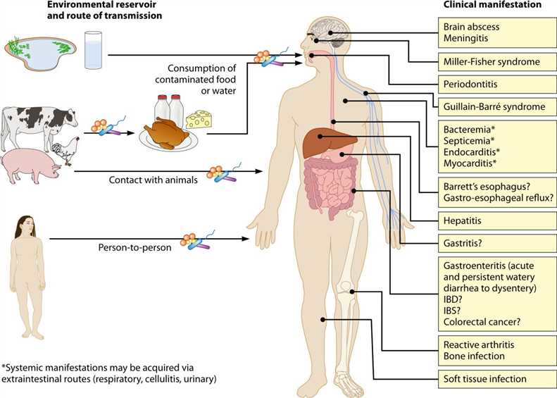 Fig. 1 Environmental reservoirs, routes of transmission, and clinical manifestations associated with Campylobacter species (Kaakoush NO, et al. 2015)
Campylobacter is a motile, spiral, gram-negative bacterial genus. They are 0.5-5 microns long and 0.2 to 0.9 microns wide. The cells contain an outer membrane and an inner membrane, and there is periplasm between the two membranes. The outer membrane is composed of lipopolysaccharide, which embed on membrane proteins. They are antigenic with the function of invading the host. At the same time, Campylobacter is very sensitive to environmental pressure, such as heating, drying and high oxygen concentration. There are dozens of kinds of bacteria, three of which are the main sources of human infection: Campylobacter jejuni (C. jejuni), Campylobacter coli (C. coli) and Campylobacter lari (C. lari). C. Jejuni is the most common implicated species. C. jejuni was reported in 1931 by Jones et al., which is commonly associated with poultry. It is considered to be one of the most important species of Campylobacter which is responsible for 80 to 85% of all enteric Campylobacter infections. Different from C. jejuni, pigs are the main source of C. Coli, but their infection symptoms are similar. C. Coli causes 10-15% of Campylobacter infections. C. Lari was first classified by Benjamin et al. in 1984, and then divided into two subspecies, C. lari concheus and C. lari lari, by Debruyne et al. in 2009.
Table 1 Species within the genus Campylobacter and their clinical relevance to humans (Kaakoush NO, et al. 2015)
| Campylobacter species | Clinical manifestations |
| C. coli | Established pathogen in gastroenteritis; also found in blood, meningitis, and acute cholecystitis |
| C. concisus | Emerging pathogen associated with gastroenteritis and IBD (Crohn's disease and ulcerative colitis); also found in Barrett's esophagitis, blood, and brain abscess |
| C. curvus | Found in gastroenteritis, ulcerative colitis, Barrett's esophagitis, blood, liver, and bronchial abscesses |
| C. fetus | Associated with bacteremia; also found in gastroenteritis, brain abscesses, epidural abscess aspirate, cerebrospinal fluid, cellulitis, endocarditis, mycotic aneurysm of the abdominal aorta, and peritonitis |
| C. gracilis | Potential periodontal pathogen; also found in IBD, head and neck infection, and brain abscess |
| C. hominis | Found in blood and IBD (possibly a commensal in the intestine) |
| C. helveticus | Found in gastroenteritis |
| C. hyointestinalis | Found in gastroenteritis and blood |
| C. insulaenigrae | Found in gastroenteritis and blood |
| C. jejuni | Established pathogen in gastroenteritis and possible predisposing agent in IBD, postinfectious IBS, and celiac disease; infection may result in sequelae in the forms of Guillain-Barré syndrome, Miller Fisher syndrome, Bell's palsy (unilateral facial paralysis), and reactive arthritis; found in IBD, blood, myocarditis, meningitis, acute cholecystitis, urinary tract infection, and acute febrile illnesses associated with leukopenia or thrombocytopenia |
| C. lari | Associated with gastroenteritis; also found in blood |
| C. mucosalis | Found in gastroenteritis |
| C. rectus | Putative periodontal pathogen; also found in gastroenteritis, IBD, vertebral abscess, blood, necrotizing soft tissue infection, and pus C. showae Found in IBD, intraorbital abscess, and blood |
| C. showae | Found in IBD, intraorbital abscess, and blood |
| C. sputorum | Found in gastroenteritis, axillary abscess, and blood |
| C. upsaliensis | Emerging pathogen in gastroenteritis; also found in breast abscess, blood, and placenta |
| C. ureolyticus | Associated with gastroenteritis and IBD; also found in oral, perianal, and soft tissue abscesses, soft tissue or bone infections, and ulcers or gangrenous lesions of the lower limb |
The study on the pathogenesis of C. jejuni shows that bacteria first penetrate gastrointestinal mucus, adhere to intestinal cells, and then cause diarrhea through toxin release. It will release several different toxins, mainly enterotoxin and cytotoxin. These toxins vary from strain to strain and are related to the severity of enteritis. During infection, all immunoglobulin levels rise. Among them, IgA is the most important due to its penetrability through the intestinal wall. IgA is able to immobilize organisms, enable them to aggregate and activate complements, and provide short-term immunity against infected strains.
While gastroenteritis is a major clinical condition resulting from Campylobacter infection, these organisms have also been associated with a range of other serious conditions within the gastrointestinal tract, including IBD, esophageal diseases, periodontitis, functional gastrointestinal disorders, celiac disease, cholecystitis, and colon cancer. In addition to gastrointestinal infection, Campylobacter species also cause a range of clinical manifestations in other parts of the body, as either a local isolated infection, a systemic manifestation after an episode of enteritis, or a postinfectious immune disorder. These manifestations include Guillain-Barré syndrome, Miller Fisher syndrome, brain abscesses and meningitis, bacteremia, sepsis, endocarditis and myocarditis, reactive arthritis, and clinical manifestations that result in complications in the reproductive tract.The most important post-infectious manifestation is Guillain-Barré syndrome, 30% - 50% of which is caused by Campylobacter infection. This syndrome is an acute demyelinating disease that affects peripheral neurons and is characterized by ascending paralysis.
Campylobacter infection is diagnosed when a laboratory test detects Campylobacter bacteria in stool, body tissue, or fluids. The test methods include culture, enzyme immunoassay (EIA), or PCR. A commercial probe assay directed against Campylobacter RNA detects C. jejuni subsp. jejuni, C. jejuni subsp. doylei, C. coli and C. lari and is highly sensitive; however, cross hybridisation with C. hyointestinalis has been noted for some isolates. Serological assay can be used to determine the antecedent cause of the Guillin-Barré syndrome as well as the role of Campylobacter in cases of reactive arthritis. Immunoglobulin G (IgG), IgM, and IgA levels in serum rise in response of infection. IgA levels in serum and faeces increase during the first few weeks of infection and then fall rapidly. Serum antibody assays vary in both sensitivity and specificity for detecting Campylobacter infection, and test performance appears to be population dependent.
Most clinical cases of campylobacter enteritis are mild or self-limited, and antibiotics are generally used in a few severe or recurrent cases. Macrolides are the first choice of drugs, and the resistance rate to erythromycin is relatively low. Severe systemic infections can be treated with aminoglycoside drugs such as gentamicin. Fluoroquinolones are effective drugs for the treatment of acute bacterial diarrhea, which can fight against most intestinal pathogens. Ciprofloxacin is widely used as a preventive drug for travelling. However, the emergence of resistance to these drugs makes their efficacy less certain. It is reported that the resistance of Campylobacter to macrolides and fluoroquinolones has increased significantly since the 1990s.
References
Loading ......
