
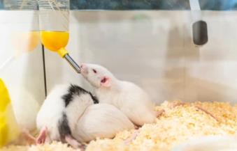
 Fig 1. Oral Administration Mice
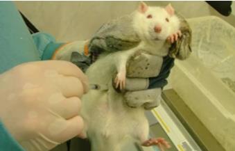 Fig 2. Intraperitoneal Injuction Mice
Inquiry Basket
 Loading ......
Cell proliferation is a fundamental process in biology, playing a crucial role in development, tissue repair, and disease progression. Labeling and tracking dividing cells is essential for understanding these processes and identifying potential therapeutic targets. One widely used technique for labeling and tracking proliferating cells is BrdU staining.
BrdU staining is based on the incorporation of 5-bromo-2'-deoxyuridine (BrdU) into the DNA of dividing cells. BrdU is a derivative of uridine that, when added to cultured cells, incorporates substituted thymidine into the DNA of cells in the S phase. During the staining process, the DNA must be degraded by DNase I, which is critical to the staining success. Staining was then performed with an anti-BrdU antibody to identify the incorporation of BrdU into proliferating S-phase cells. BrdU staining is an advantageous alternative for customers assessing cell proliferation using traditional [3H]thymidine incorporation methods. BrdU staining can help customers identify cells that have entered the S-proliferative phase triggered by stimulators or proliferation-inducing treatments.
The BrdU staining has a wide range of applications in cell biology, developmental biology, and cancer research. One common use is to study cell proliferation in vitro, by labeling cells with BrdU and monitoring the incorporation of BrdU into DNA over time. This can provide insights into the kinetics of cell proliferation, such as the length of the cell cycle and the rate of cell division. In cancer research, BrdU staining can be used to study the proliferation of tumor cells and to identify potential therapeutic targets.
BrdU staining can also be used to study cell proliferation in vivo, by injecting BrdU into animals and tracking its incorporation into DNA in various tissues. This can be used to study tissue-specific proliferation rates, as well as the effects of various treatments or genetic manipulations on cell proliferation.
1) Dissolve 3 mg of BrdU in 1 mL of water to prepare a 10 mM BrdU stock solution.
2) Dilute 10 mM BrdU stock solution in cell culture medium to prepare 10 μM BrdU labeling solution.
3) Filter 10 µM BrdU labeling solution through a 0.2 µm filter (sterile conditions).
4) Replace existing medium in cells with 10 μM labeling solution.
5) Incubate cells in the BrdU labeling solution in a CO2 incubator at 37ºC for 1-24 hours.
Note: BrdU incubation time depends on cell division rate. The exact time needed to achieve the best signal-to-noise ratio should be optimized.
6) Remove the BrdU labeling solution from cells.
7) Wash twice in PBS for approximately 5 seconds each.
8) Wash three times in PBS for two minutes each.
9) Fix and permeabilize cells according to standard immunocytochemistry (ICC)/immunofluorescence (IF) protocols. See below for DNA hydrolysis steps prior to immunostaining.
Oral administration and intraperitoneal injection are common methods of labeling cells with BrdU in vivo.
| Oral Administration | Intraperitoneal Injection |
2) After BrdU treatment, animals can be sacrificed according to standard protocols. 3) Fix and process tissue according to standard IHC protocols. See below for DNA hydrolysis steps prior to immunostaining. Note: The exact dose should be optimized for individual experimental conditions. |
2) For mice, BrdU solutions are typically injected to 100 mg/kg. 3) After BrdU treatment, animals can be sacrificed according to standard protocols. 4) Fix and process tissue according to standard IHC protocols. See below for DNA hydrolysis steps prior to immunostaining. Note: The exact treatment time and dose should be optimized according to the tissue of interest. |
| Cells | Tissues Sections |
| 1) Incubate cells in 1-2.5 M HCl for 10 minutes to 1 hour at room temperature. Note: The exact HCl concentration and incubation time/temperature should be optimized according to the specific experiment. 2) (Optional) Remove HCl and neutralize with 0.1 M sodium borate buffer (pH 8.5) for 30 minutes at room temperature. 3) Wash 3 times in PBS for approximately 5 seconds each. 4) Proceed with immunostaining according to ICC protocols. | 1) Incubate sections in 1-2 M HCl for 30 min to 1 h. Note: The exact HCl concentration and incubation time/temperature should be optimized according to the specific experiment. 2) (Optional) Neutralize sections by incubating them in 0.1 M sodium borate buffer (pH 8.5) for 10 minutes at room temperature. 3) Wash 3 times in PBS for approximately 5 seconds each. 4) Proceed with immunostaining according to IHC protocols. |
In addition to BrdU staining, co-staining with other antibodies (e.g., Ki67, NeuN, Doublecortin) can provide valuable information about the localization and function of proliferating cells. For example, NeuN, which is a marker of mature neurons, can be used in conjunction with BrdU staining to identify newly differentiated neurons.
Creative Diagnostics is a leading provider of products and services for cell biology and cancer research, including a wide range of reagents and kits for BrdU staining. Our BrdU Cell Proliferation Assay Kit provides a simple, sensitive, and reliable method for detecting cell proliferation based on BrdU incorporation. The kit includes all the necessary reagents for labeling, fixing, and staining cells, as well as a detailed protocol and troubleshooting guide.
Creative Diagnostics also offers a wide range of Anti-BrdU Antibodies, including monoclonal and polyclonal antibodies validated for use in various applications, such as immunofluorescence, immunohistochemistry, and flow cytometry. Our products are of high quality and have been extensively tested in various applications, ensuring reliable and reproducible results.
| Target | Cat. No. | Product Name | Size | Application | Detection Sample | |
| BrdU | DEIA8695 | Cellular DNA Fragmentation ELISA Kit | 96T | Qualitative | cytoplasm, culture supernatant | Inquiry |
| DEIA8698 | Cell Proliferation ELISA Kit, BrdU (chemiluminescent) | 96T | Qualitative | cells | Inquiry | |
| DEIA8699 | Cell Proliferation ELISA Kit, BrdU (colorimetric) | 96T | Qualitative | replicating (cycling) cells | Inquiry |
Loading ......
