
Inquiry Basket
 Loading ......
Antibody-drug conjugates (ADCs) are an emerging class of targeted therapies for the treatment of cancer that aim to directly deliver small molecule chemotherapy and kill tumor cells while sparing healthy cells. Since the successful approval of MYLOTARG in 2000, antibody drug conjugates (ADCs) have become a key category of targeted cancer therapy. They mainly contain three parts: monoclonal antibodies, linkers and toxins. Monoclonal antibodies are the key to determining the efficacy of ADC; the linker determines the systemic toxicity and clinical efficacy of ADC drugs; toxins are the key factor that determines the lethality of ADC drugs. Although there are currently more than 130 types of toxins used in announced ADC drugs, there are only 6-8 toxins used in ADC drugs that are in clinical trials or have been marketed. According to the mode of action, they can be roughly divided into three categories: tubulin inhibitors, DNA synthesis inhibitors and topoisomerase inhibitors. Among them, ADC drugs using MMAE in tubulin inhibitors as toxin molecules have been approved the most. Today we focus on the definition of MMAE and its application and mechanism of action in ADC.
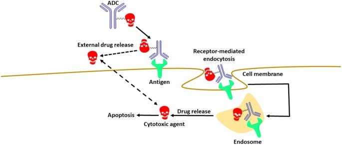 Figure 1. A schematic view of the mode by which ADCs function. (Source: Johansson MP, et al., 2017)
Before we start introducing MMAE, let’s first understand the development history of toxins in ADC drugs. The concept of ADC drugs was first proposed by German Nobel Prize winner Paul Ehrlich in 1913. But ADC drug development began in 1975, because that was when hybridoma technology began to be used to produce monoclonal antibodies. After nearly 50 years of research and development, ADC drugs have undergone three iterative updates. Among these three generations of ADC drugs, the toxins involved also have their own characteristics:
The first generation of ADC drugs mainly uses traditional chemotherapy drugs as toxins, such as calikamycin. However, because traditional chemotherapy drugs have weak cytotoxicity and specificity against cancer cells, and are not easy to accumulate in target cells, the efficacy of this generation of ADC is even worse than that of chemical drugs alone. In addition, the first-generation ADC drugs also have shortcomings such as unstable linkers, highly hydrophobic cytotoxicity, and poor conjugation technology.
Compared with the first-generation ADC drugs, the second-generation ADC drugs have comprehensive optimization of the three components of antibody, linker and cytotoxin. As far as cytotoxins are concerned, the toxins used in second-generation ADC drugs are mainly tubulin inhibitors, such as MMAE, MMAF, etc. This is a new class of highly cytotoxic compounds that are 100 to 1,000 times more toxic to cancer cells than traditional chemotherapy drugs used in first-generation ADCs. But it is worth noting that these toxins often produce intolerable side effects when used as single drugs to kill tumors, so they are not approved as single drugs for cancer treatment. However, this class of toxins is ideal for toxin selection in ADC drug development. Because tumor cells that divide more rapidly are more sensitive to tubulin inhibitors than normal cells. But this type of toxin also has a drawback, that is, they are more effective at killing dividing tumor cells than static cancer cells.
In the development process of third-generation ADC drugs, in order to avoid this limitation, most drugs choose DNA damaging agents that can target the entire cell cycle as conjugated toxins, such as enediyne, topoisomerase I inhibitors and pyrrolo Benzodiazepines. DNA-damaging agents can kill tumor cells by disrupting DNA structure through double-strand breaks, alkylation, chimerization, and cross-linking. In addition, the third generation of ADC drugs has also made significant progress in conjugation technology and antibody selection.
Although ADC drugs have been iterated three times, the selection of ADC drug-conjugated toxins still has clinical limitations, such as severe side effects and drug resistance. Among the 15 ADC drugs currently approved, 5 ADC drugs use MMAE as a toxin molecule.
Monomethyl auristatin E (MMAE) is a cytotoxin that can kill cancer cells. MMAE is an analogue of Dolastatin 10. Microtubules are composed of α-tubulin and β-tubulin and are the main components of the cytoskeleton. Tubulin inhibitors kill tumor cells by destroying their cytoskeletal structure and interfering with mitosis. Dolastatin 10 is isolated from the Indian Dolabella Auricularia. It is a microtubule inhibitor and has extremely strong cytotoxicity. In addition to MMAE, the synthetic Dolastatin 10 analogues also include monomethyl auristatin F (MMAF), both of which are widely used as ADC payload components, that is, ADC toxins. MMAE contains four amino acids: monomethylvaline (MeVal), valine (Val), dolaysoleucine (Dil), and dolaproline (Dap), as well as the carboxyl-terminal amine norephedrine. In MMAF, the C-terminus of monomethylvaline is replaced by phenylalanine, and its cellular activity is significantly reduced. MMAE is essentially demethylated auristatin E, that is, the N-terminal amino group has only one methyl substituent instead of two methyl substituents like auristatin E itself. MMAE is a synthetic anti-tumor drug that is 100-1000 times more potent than doxorubicin. Due to its toxicity, MMAE cannot be used as a drug by itself. But after it is connected to a monoclonal antibody, because the monoclonal antibody can recognize specific markers in cancer cells, MMAE can be guided to specific cancer cells.
Since ADC toxins are highly cytotoxic, PK studies of ADC payloads are important to understand the behavior of the payload. However, due to the high toxicity of MMAE itself, such studies are not allowed in humans. Therefore, it is very important to detect MMAE levels preclinically (such as measuring the concentration of ADC and conjugated MMAE). The ADC ELISA developed using anti-MMAE antibodies is used to quantitatively determine the level of anti-MMAE-conjugates in test samples. It can be used for PK detection and DAR value analysis to accelerate the development process of ADC drugs.
Reference
| Target | Cat. No. | Product Name | Host | Isotype | Application | |
| MMAE | CABT-B8992 | Anti-MMAE monoclonal antibody, clone 3F3 | Mouse | IgG1 | ELISA | Inquiry |
| CABT-B8993 | Anti-MMAE monoclonal antibody, clone 3F3 [Biotin] | Mouse | IgG1 | ELISA | Inquiry | |
| CABT-L3100 | Rabbit Anti-vc-PAB-MMAE polyclonal antibody | Rabbit | IgG | ELISA | Inquiry | |
| CABT-L3101 | Mouse Anti-vc-PAB-MMAE monoclonal antibody, clone C22G22 | Mouse | IgG | ELISA | Inquiry | |
| CABT-L3103 | Mouse Anti-MMAE / F monoclonal antibody, clone C23B3 | Mouse | IgG | ELISA | Inquiry | |
| CABT-ZB1143 | Mouse Anti-MMAE monoclonal antibody, clone 3F3 [HRP] | Mouse | IgG1 | ELISA | Inquiry | |
| CABT-ZB1144 | Mouse Anti-MMAE monoclonal antibody, clone 3F3 [AF647] | Mouse | IgG1 | ELISA | Inquiry | |
| CABT-B8997 | Anti-MMAE polyclonal antibody | Mouse | IgG | ELISA | Inquiry |
| Target | Cat. No. | Product Name | Size | Species Reactivity | Application | Detection Sample | |
| MMAE | DEIABL312 | MMAE ADC EIA Kit | 96T | Quantitative | tissue cell cultures, serum | Inquiry | |
| DEIABL314 | Intact MMAE ADC ELISA Kit | 96T | Human | Quantitative | Serum | Inquiry |
Loading ......
