
Inquiry Basket
 Loading ......
CD20 is encoded by the MS4A1 gene. CD20 is a non-glycosylated membrane protein of molecular weight ~33–37 kDa. CD20 is a member of the membrane spanning 4-domains protein family (MS4A). There are 18 members of MS4A family in humans. MS4A1 has structural homology to the other members of the MS4A gene family. All the genes are physically linked on the human chromosome in the 11q12 region, except MS4A4. The MS4A1 gene spans about 16 kb and contains 8 exons. There are several CD20 mRNA transcripts, the most abundant is a 2.8 kb transcript containing all 8 exons. The second most common variant is a 263 bp transcript where exon II is skipped. A third isoform uses a 3' splice site in the first intron to generate a shorter 3.5 kb transcript, but all three transcripts use the same translational start site in exon III to produce the full length CD20 protein. There are a few examples of abnormal splicing variants in malignant B cells, in which truncated forms of CD20 are produced. These have been shown to be associated with defective binding of anti-CD20 mAbs.
 Figure 1. A schematic of the proximal region of MS4A1 promoter with transcription factor binding sites
(Source: Pavlasova G, et al. 2020)
CD20 is a protein with 4 hydrophobic transmembrane domains, one intracellular tail and two extracellular loops of unequal length. The N- and C-termini are cytoplasmic. The protein is phosphorylatable, and at least three homologous subtypes have been described (33, 35, 37 kDa). Phosphorylation is greater on proliferating malignant B cells compared to resting B cells. CD20 exists on the cell membrane as homodimers or tetramers. It is also involved in supramolecular complex formation with a number of other molecules (CD53, CD81, CD82, MHCII, CD40, BCR, CBP, Src family kinases, etc.) and is involved in signal transduction. The degree of CD20 expression varies widely on B-cell malignancies and is believed to be the lowest in patients with CLL and highest in DLBCL and hairy cell leukemia; within CLL, mutated IGHV patients express more CD20 than unmutated IGHV CLL. There are some data that high CD20 expression is associated with longer overall survival following treatment with rituximab, though this is controversial. CD20 expression is also highly heterogeneous between individual patients and among different cellular subpopulations within a clone.
The function of CD20 on normal and malignant B cells is not yet fully understood. There is some evidence that CD20 is intimately linked to the B-cell receptor (BCR) signalling pathway, as well as to the microenvironmental interactions. The few mutations in MS4A1 that cause a complete absence of the cell surface protein CD20 result in decreased numbers of memory B cells in vivo, decreased immunoglobulin IgG levels, and an otherwise clinically impaired response to T-independent antigens (for example, pneumococcal polysaccharides) with a normal response to T-dependent antigens (for example, tetanus toxoid). CD20 can also be found in "lipid raft" microdomains and directly co-localize with the BCR. After BCR activation, CD20 is highly phosphorylated, and displays calcium channel-like properties, suggesting a role in calcium influx and downstream signaling. siRNA-mediated downregulation of CD20 on human B-cell lines strongly inhibits BCR-mediated calcium influx, and direct anti-CD20 cross-linking is also able to induce calcium signalling and downstream gene expression responses that resemble those upon BCR cross-linking. CD20 is upregulated on cells that exit lymph nodes and enter the peripheral blood of CLL patients through the CXCR4-SDF1 pathway, and the microenvironment-associated factors IL-4, TNFα and IFNα can also induce CD20 expression in vitro. This might at least partly explain the suboptimal activity of the BTK-inhibitor ibrutinib combined with rituximab (anti-CD20): ibrutinib blocks both BCR-signalling and the CXCR4-pathway, causing downregulation of CD20 and thereby attenuating its presence as a target for antibody action. In contrast, obinutuzumab requires lower CD20 levels and shows better combination efficacy. Additionally, CD20-deficient mice exhibit a 20–30% reduction in surface IgM levels and impaired BCR/CD19-dependent calcium mobilization, which some studies attribute to impaired calcium transport rather than storage release issues. Silencing CD20 in malignant B cells affects the phosphorylation of BCR-associated kinases (LYN, SYK) and downstream proteins (GAB1, ERK), suggesting that CD20 may participate in both "initiating and sustaining" BCR signaling or act as a "basal regulator."
B-cell development proceeds through antigen-independent maturation in the bone marrow from hematopoietic stem cells to pro-B cells, then into immature B cells, followed by antigen-dependent maturation in peripheral lymphoid tissues. Pro-B cells (CD19+, CD20+) develop from precursor B cells (CD19−, CD20−) and then further mature to become immature B cells in the bone marrow where they begin to express IgM. After antigen-specific activation and co-stimulatory factors, these cells further mature to become mature B cells. Activated (immunoglobulin isotype switched) cells will leave the germinal center to go to sites like the bone marrow, brain, intestine, spleen, and tonsils to differentiate into memory B cells (low CD27 expression) or plasmablasts (early/high CD27 and CD40L+, late/CD27+ and CD38+). Chemokines specific to the site of travel (CXCL12, CCL25, and CCL28, among others) guide these cells to differentiate into antibody-secreting plasma cells. At these locations, B cells may also serve as antigen-presenting cells, or produce pro-inflammatory cytokines to promote the inflammatory response.
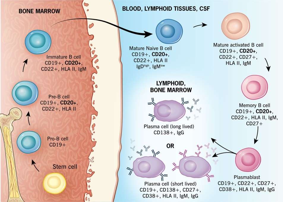 Figure 2. B-cell maturation
(Source: Carlson AK, et al. 2024)
Different authors have found B-cell and T-cell infiltrates to be co-localized in active lesions and perivenular spaces in progressive MS patients, and have also noted the presence of abundant plasma cells and CD20+ B cells. In addition, deep sequencing of the IgG heavy chain variable region genes has revealed small clonally related B-cell populations, implying peripheral B-cell activation and the notion that B-cell maturation and immune responses take place in parallel in the periphery and CNS. Activated B cells typically interact with T-helper (Th) cells in the germinal centers, differentiating into memory B cells while inducing effector activation of Th cells. It is believed that in MS patients, peripheral B cells escape the control of functionally impaired regulatory T cells.
CD20 has been proposed to work as an ion channel, or to indirectly modulate calcium release triggered by the B-cell antigen receptor. Depletion of B cells with anti-CD20 monoclonal antibodies affects not only circulating B cells, but also a fraction of CD3+, CD4+, and CD8+ T cells that express CD20. The B-cell depletion results from a combination of different mechanisms, including apoptosis, complement-dependent cytotoxicity and ADCC. Stem cells, pro-B cells, plasmablasts and antibody-secreting plasma cells are not targeted, as they do not express CD20. Four anti-CD20 monoclonal antibodies are currently approved for the treatment of multiple sclerosis: rituximab, ocrelizumab, ofatumumab and ublituximab. The four drugs have different molecular structure, epitope targeted, and pharmacologic properties, including route of administration and dosing regimen, as well as mechanism of B-cell depletion, patterns of B-cell repopulation, risk profile and immunogenicity. Their capacity to mediate CDC and ADCC after binding also differs.
Table 1. Pre-treatment testing and safety monitoring for anti-CD20 monoclonal antibody therapy
| Contraindications | Active hepatitis B infection History of life-threatening infusion reaction to medication |
| Recommended pretreatment testing | Complete blood count with differential Comprehensive metabolic panel Quantiferon/tuberculosis screen Hepatitis panel Varicella zoster virus IgG (to ensure immunity) Quantitative Ig levels Urine or serum beta human chorionic gonadotropin (where appropriate) Brain MRI All necessary vaccinations > 4 weeks prior to first dose |
| Recommended monitoring schedule | Complete blood count with differential (every 6 months) Comprehensive metabolic panel (every 6 months) CD19 count (every 6 months) Quantitative Ig levels (every 6 months) Monitor for recurrent or serious infection Monitor for malignancy/ensure all age-appropriate cancer screening up to date |
| Pregnancy and family planning | Recommend contraception during treatment and for 6 months after |
(Source: Carlson AK, et al. 2024)
B cell depletion achieved by administration of mAbs can lead to infusion-related reactions (IRRs) and injection-related reactions. A number of mechanisms have been proposed, of which complement-dependent cytotoxicity is a relatively potent effector. Compared with rituximab and ofatumumab, ocrelizumab and ublituximab have a lower incidence of infusion or injection-related reactions likely due to their weak CDC activity. B cells have a protective role in infection and tumor surveillance, and this may occur by both antibody-dependent and antibody-independent pathways. Since plasma cells do not express CD20, long-term administration of anti-CD20 can lead to hypogammaglobulinemia. In this scenario, several management approaches are possible, including dose reduction or prolongation of mAb dosing interval, intravenous or subcutaneous supplementation of immunoglobulin, or discontinuation and switch to another DMT.
Anti-CD20 therapies have also been shown to affect T cell populations. It was found that anti-CD20 therapy not only directly depletes CD20+ T cells, but can also indirectly modulate other T cell populations by disrupting B:T cell interactions. In hematological studies, the number of CD20+ T cells was observed to rapidly decline in MS patients following treatment with ocrelizumab. CD3+CD20+ T cells were demonstrated to be nearly completely eradicated by 12 weeks of treatment with rituximab. In a different study, ofatumumab was also demonstrated to have the ability to modulate regulatory T cells with resultant decreases in non-suppressive regulatory T cells, increases in naive regulatory T cells and a reduced ratio of circulating T follicular helper cells to circulating T follicular regulatory cells. Other studies have also shown similar changes to T cell populations following ocrelizumab treatment. While the clinical relevance of CD20+ T cell depletion remains to be determined, it has been proposed as one possible mechanism of action of anti-CD20 monoclonal antibody therapies.
References
| Target | Cat. No. | Product Name | Size | Species | Application | Detection Sample | |
| CD20 | DEIA-LL086 | Mouse CD20 ELISA Kit | 96T | Mouse | Quantitative | Serum, plasma, tissue homogenates and other biological fluids | Inquiry |
| Target | Cat. No. | Product Name | Expression System | Tag/Conjugate | Application | |
| CD20 | DAG-CL002 | Human CD20 (aa 213 - 297) [His] | HEK293 | His | N/A | Inquiry |
| CD20 | DAG-WT1241 | Recombinant Human CD20 VLP | HEK293 cells | N/A | ELISA, SPR | Inquiry |
| CD20 | DAG-WT1245 | Biotinylated Recombinant Human CD20 VLP | HEK293 cells | N/A | ELISA, SPR | Inquiry |
Loading ......
