Case Studies for Spatial Proteomics
Imaging Analysis of Spatial Proteomics
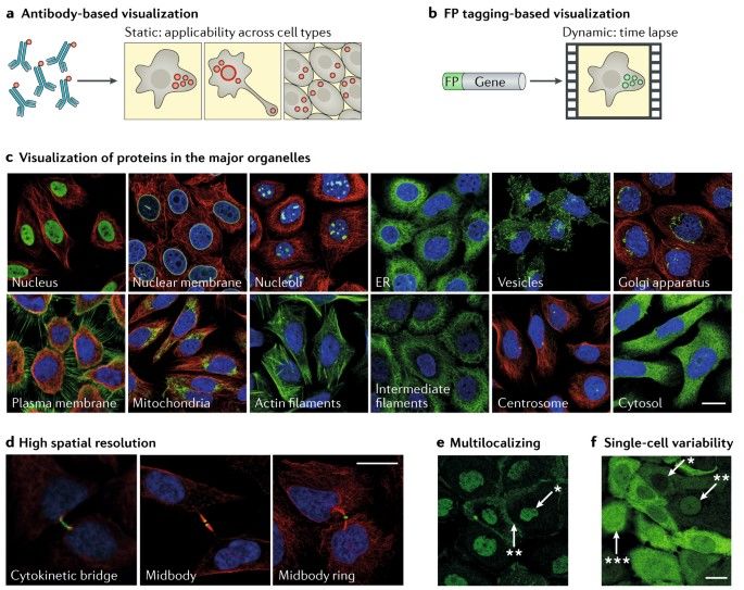 Different approaches to imaging-based spatial proteomics. (Lundberg E, et al.; 2019)
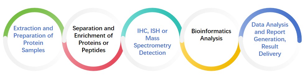
With our team's extensive expertise in Spatial Proteomics analysis we deliver premium Spatial Proteomics services which cover quantitative proteomics alongside protein qualitative and modified proteomics analysis. Our technology platform delivers complete proteomics services by integrating strict quality control measures with professional analytical procedures.
Spatial proteomics explores where proteins are located within cells and tissues and what roles they perform in these environments. High-resolution imaging technology paired with mass spectrometry analysis and bioinformatics methods enables researchers to examine protein localization and subcellular distribution as well as protein interactions on both cellular and biological levels. Spatial proteomics enables scientists to acquire detailed protein expression profiles of individual cells and tissue regions which reveal proteins' specific spatial expressions.
Spatial proteomics employs key technologies such as immunofluorescence labeling and mass spectrometry imaging technology. These technologies enable scientists to determine cellular protein locations, understand protein interactions with DNA, RNA and other molecules and finally uncover organisms' complex regulatory networks. The application of spatial proteomics extends to functional regional proteome analysis of complex tissues and heterogeneous proteome analysis of cancer tissues which enables researchers to understand neuronal functions and activation states while identifying abnormal activation pathways and potential therapeutic targets.
Spatial proteomics primarily uses three technical methods which are imaging mass spectrometry (MALDI-MS imaging), in situ sequencing (CITE-seq) and deep visualization proteomics (DVP). Through laser capture microdissection and mass spectrometry analysis imaging mass spectrometry technology generates high-resolution protein localization information by mapping protein spatial distributions on tissue sections. In situ sequencing technology combines oligonucleotide spatial tags with antibodies to perform in situ protein capture and sequencing for targeted spatial distribution detection of specific proteins. Deep visual proteomics (DVP) can accurately map the spatial distribution of proteins through laser microdissection and mass spectrometry analysis combined with single-cell samples. These technical methods provide important tools for studying the spatial distribution of proteins in cells and tissues.
| Multiplex immunofluorescence (mIHC/mIF) | Through cyclic staining techniques (such as CycIF and IBEX), co-localization analysis of 30-100 proteins in a single sample can be achieved, supporting targeted detection of cell membrane, cytoplasmic and nuclear proteins. |
| CODEX Technology | Using DNA barcodes to label antibodies, sequencing and decoding can achieve simultaneous imaging of more than 100 proteins, which is suitable for analyzing the immune microenvironment (such as the distribution of tumor-infiltrating lymphocytes) |
| Imaging Mass Cytometry (IMC) | Combining metal chelating antibodies with mass spectrometry detection, it provides subcellular resolution protein spatial distribution maps and supports multi-omics joint analysis (such as RNA-protein co-localization) |
| Laser microdissection (LCM) combined with mass spectrometry | By capturing tissue micro-regions (such as tumor core areas and perivascular areas) with high precision laser, proteomic analysis from single cell to multi-cell level can be achieved while retaining spatial background information. |
| Deep Visual Proteomics (DVP) | The spatial distribution of proteins, metabolites and lipids can be directly detected without labeling, covering the unbiased analysis of the entire proteome, and is suitable for drug metabolism and tissue heterogeneity studies. |
| Mass spectrometry imaging (MSI) | Integrating microdissection with ultra-high sensitivity mass spectrometry breaks through the limitations of antibodies and provides proteomic data at single-cell resolution. |
| Protein-transcriptome Joint Analysis | By combining viewRNA with immunofluorescence, the co-localization of protein and RNA can be simultaneously detected to reveal the post-translational regulatory mechanism. |
| Proteome-Metabolome Integration | Mass spectrometry imaging technology is used to construct a spatial interaction network of metabolic enzymes, substrates and products (such as the spatial dynamics of tumor glycolysis pathways). |
| Metallosome-Proteome Association | Analyze the distribution of metal elements (such as iron and zinc) and their functional association with metal-binding proteins. |
| Spatial Heterogeneity Analysis | Map protein abundance gradients, subcellular localization, and spatial boundaries of functional modules in complex samples such as tumors and neural tissues (e.g., differences in immune checkpoint proteins between the edge and core of a tumor). |
| Identify protein expression signatures of rare cell populations (e.g., circulating tumor cells, stem cell niches). | |
| Dynamic Process Tracking | Temporal changes in protein localization during embryonic development, tissue repair, etc. (e.g., spatial activation patterns of key signaling pathways in cardiac development). |
| Evaluation of protein spatial redistribution after drug treatment (e.g., the effect of chemotherapy drugs on PD-L1 expression in the tumor microenvironment) |
Imaging Analysis of Spatial Proteomics
Different approaches to imaging-based spatial proteomics. (Lundberg E, et al.; 2019)
High spatial resolution: Spatial proteomics technology can locate and quantify proteins at the fine scale of cells, tissues and even sub-cells. This technology can provide spatial information different from traditional proteomics, helping researchers understand the distribution of proteins in different biological tissues and their biological functions.
Multi-level analysis capabilities: Our spatial proteomics can simultaneously analyze different types of proteins, including membrane proteins, cytoplasmic proteins and nuclear proteins, and can handle complex samples such as tissue sections and single-cell samples.
High sensitivity and high throughput: Our services should be able to detect low-abundance proteins and perform large-scale spatial protein quantitative analysis. Using highly sensitive detection methods and advanced mass spectrometry analysis technology, thousands of proteins can be analyzed simultaneously.
Accurate spatial positioning: The technology should be able to accurately mark the spatial position of proteins in tissue sections, helping researchers reveal the distribution and dynamic changes of proteins in different tissues, cell types or cell substructures.
Strong data analysis capabilities: Our spatial proteomics services not only provide raw data, but also perform high-level data analysis to help customers extract meaningful information from complex data and draw scientific conclusions. Including multidimensional data analysis, pattern recognition and spatial association analysis.
Highly customized services: Ability to provide personalized solutions based on different research needs. Researchers receive customized proteomics analysis services when studying particular diseases or analyzing different cell types and biomarkers.
Technological innovation: The field of spatial proteomics services integrates leading-edge technologies like laser capture microdissection (LCM) and in situ mass spectrometry imaging because these tools deliver superior analytical precision and diverse application possibilities.
Data and experimental design support: Top-notch service providers extend data interpretation and experimental design assistance to maintain scientific validity in research plans and the reliability of collected data while helping customers achieve better experimental outcomes.
Spatial proteomics maps protein distributions in cells and tissues while maintaining spatial context which allows researchers to investigate biological system interactions. Nature Methods recognized spatial proteomics as 2024 Method of the Year because of its groundbreaking potential to reveal cellular connectivity and disease mechanisms.
This technology connects single-cell genomics with spatial transcriptomics and functional phenotyping to offer integrative analysis. The validation of biomarkers detected by spatial transcriptomics takes place at the protein level through Akoya's PhenoCycler-Fusion (supporting 20+ markers) or PhenoImagerHT (capable of 9-plex fluorescence) which facilitates comprehensive multi-omics analysis.
Mass spectrometry-based: High-resolution platforms like Orbitrap™ Astral™ provide deep proteome coverage.
Imaging-based: Fluorescence imaging (e.g., Akoya's STEP Kit) uses barcoded antibodies and cyclic staining to map 100+ proteins at single-cell resolution.
AI-driven: Artificial intelligence combines spatial transcriptomics with histology data to accurately determine protein locations.
Tumor microenvironment analysis: The technique reveals key immune cell interactions and therapeutic targets within lung cancer and melanoma tissues.
Neurology and development: Examines protein network structures within brain tissues and stem cells.
Clinical translation: The clinical translation application enables biomarker validation as well as drug efficacy assessment and prognostic prediction.
Challenges: Multiplexing capacity remains restricted while costs stay high and data integration presents significant complexity challenges.
Future trends: Future developments will see improved AI tools alongside scalable operational procedures and CRISPR-based protein editing integration. The focus of publications such as Nature Biotechnology and Cell on spatial proteomics studies indicates its growing importance.
Reference