Laser Confocal Chip Scanner
Biochips work on analytic hybridisation. The basic solution is to stain the sample to be examined, then whole-body hybridise it with a known structure biochip. Cleaning and scanning take their product, which comes out as images. The biochip itself is so complex that we need to know where the hybridization signal is on the chip. The detection for weak light signal requires the presence of weak light signal detectors like photomultiplier tubes, especially for large-scale biochips because they are small, dense, have little sample, and have a low hybridisation signal. In addition, in addition to determining the distribution site of the hybridization signal, most biochips also need to determine the signal intensity at each site to determine whether it is completely hybridized or partially hybridized. Therefore, the sensitivity and linear response of the detection method are also very important. The hybridization signal detection system mainly includes three parts: hybridization signal generation, signal acquisition and transmission, and signal processing and imaging. Among them, the laser confocal biochip scanner can serve as an important technical platform for signal acquisition in this process.
What is Laser Confocal Chip Scanner?
Laser confocal biochip scanner is an advanced optoelectronic system which incorporates confocal principle, scanning and laser imaging technologies. The light source in the biochip scanner is laser due to the brightness, monochromatic, small emission angle and excellent focusing of it. The confocal biochip scanner uses conjugate focus technology to make the light source, biochip and photomultiplier tube detector in corresponding co-rotating positions. In the scanning system, the laser beam is sharply focused on the surface of the biochip through the objective lens into a diffraction-limited spot, which excites the sample containing fluorescent substances on the surface of the carrier to produce fluorescence. The fluorescence is through the optical lens and the detector lens on the confocal detection pinhole plane and it is absorbed by the photomultiplier tube near the image plane. It is detected up by the photomultiplier tube and turned into a digital signal.
Principle of Laser Confocal Chip Scanner
The laser confocal chip scanner is an advanced technology that combines the principle of confocal microscopy with the function of chip-based scanners to capture detailed high-resolution sample images. Its principle is mainly based on the confocal microscope imaging system and the confocal scanning system.
One is the confocal microscope imaging system. It consists mainly of a scanning unit, a microscope and a spectral imaging unit. In one of them, the laser in the scanning module goes into the microscope and is focused on the specimen that is being examined by the objective lens. Excited fluorescence goes into the scanning module by way of the microscope, then propagates to the dichroic mirror and through the pinhole - after being focused by the pinhole lens - into the spectral imaging module. The spectral imaging module has a collimating lens, beam splitter, focusing lens, variable slit and photomultiplier tube (PMT). Once the fluorescence arrives in the spectral imaging module, it's split into parallel light by the collimating lens, then goes into the beam splitter and focus lens. The beam splitter divides the fluorescence into a spectral band of some width and the controllable slit is selected for the spectral selection and the signal is collected by PMT.
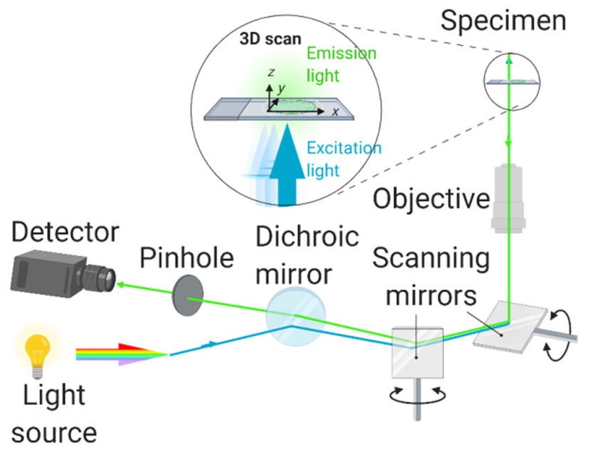 Figure 1. The basic setup of laser scanning confocal microscopy. (Sources: Xinyi Wang, et al. 2021)
It's the scanning system, a second. In laser confocal biochip scanner there are 3 kinds of confocal scanning device to realize two-dimensional scanning motion of scanning laser beam with respect to biochip in XY direction: mechanical scanning device, optical scanning device and optical-mechanical combined scanning device.
- Two-dimensional Mechanical Scanning
The device drives the working platform to perform two-dimensional mechanical movement along the axial direction and the axial direction through two motors respectively, realizing the two-dimensional scanning movement of the laser beam relative to the biochip. The scanning speed in the axial direction is faster and is driven by a linear motor. The scanning speed in the axial direction is slower and is realized by a stepper motor. The biggest advantage of this device is that the implementation method is simple and the price is low. The optical axis direction is always perpendicular to the scanning direction. The imaging quality of the microscope objective only requires points on the axis, and the aberration correction is simple. The disadvantage is that the scanning inertia is large, so the scanning speed is slow, the scanning vibration is large, and the repeatability is poor.
- Two-dimensional Optical Scanning
This device combines two scanning galvanometers in a certain way, so that the incident laser beam reaches the biochip after being reflected by the galvanometers. Through the rotation and swing of the two galvanometers, the laser beam can scan the biochip in two dimensions, and the biochip is fixed in this process. Its main advantages are that the galvanometer has a small moment of inertia, low noise, and fast scanning speed. The repetition frequency of the galvanometer can be as high as 100,000, that is, it can scan 1000 biochip images per second. The main drawback is that dual galvanometer scanning is expensive, and the imaging field of view gets larger (so the optical system has to have tiny off-axis aberrations, and it is harder to design the optical system). Meanwhile, the objective length of the microscope lens is longer, with smaller numerical aperture and poor fluorescence collection.
- Optical-mechanical 2D Scanning
This type of scanning is developed on the basis of the above two scanning devices. It fully combines the characteristics of mechanical and optical scanning system design. The laser beam scans the biochip along the direction, and the motor drives the precision guide rail to make the biochip move along the direction. The advantages of this structure are obvious. For a start, the optical distance between the objective lens and the biochip is very small, so you can create a very short focal length and the numerical aperture is very large to get a very high fluorescence capture efficiency of the biochip fluorescence analyzer. Second, the scanning field of view is linear along the optical axis, so all the objective lens needs to do is imaging the on-axis point, no aberration at the off-axis point is required, and so on. The optical system design is quite straightforward as well.
Why choose our laser confocal scanner technology platform:
- High resolution and precise imaging
- Multi-channel simultaneous detection
- High sensitivity and low background noise
- Automated data processing and analysis
- High-throughput analysis capability
- Flexible experimental customization service
- Wide range of application scenarios
- Professional support and customer service
- Stable performance and high reliability
Our Featured Services
- Protein array scanning service
- Multi-channel fluorescence imaging service
- Protein quantitative analysis service
- Protein-protein interaction analysis
- Protein epitope recognition and antibody screening service
- High-throughput protein screening service
Application of Laser Confocal Chip Scanner
- Cancer marker screening
- Drug target screening
- Antibody multi-label analysis
- Protein complex research
- Drug effect evaluation
- Immune checkpoint inhibitor research
- Virus-host cell interaction
- Antibody development and optimization
- Small molecule drug screening
- Vaccine antigen screening
- Protein expression pattern analysis
References
- Xinyi Wang, et al. Three basic types of fluorescence microscopy and recent improvement. E3S Web of Conferences. 2021, 290(5771):01031
- Sun B, et al. Microminiaturized immunoassays using quantum dots as fluorescent label by laser confocal scanning fluorescence detection. J Immunol Methods. 2001, 249(1-2):85-9.