Immune Checkpoint Molecule Detection
What are immune checkpoint molecules?
Immune checkpoints refer to a class of key molecules that regulate immune responses. The most common ones include programmed cell death protein 1 (PD-1), PD-L1, and cytotoxic T-lymphocyte antigen 4 (CTLA-4), among others. These molecules primarily regulate the activation and inhibition of immune cells to maintain immune system balance. Under normal circumstances, immune checkpoint molecules prevent overactive immune responses, protecting tissues from autoimmune attacks. However, in diseases like cancer and chronic viral infections, these molecules may be exploited by tumor cells or pathogens to evade immune surveillance.
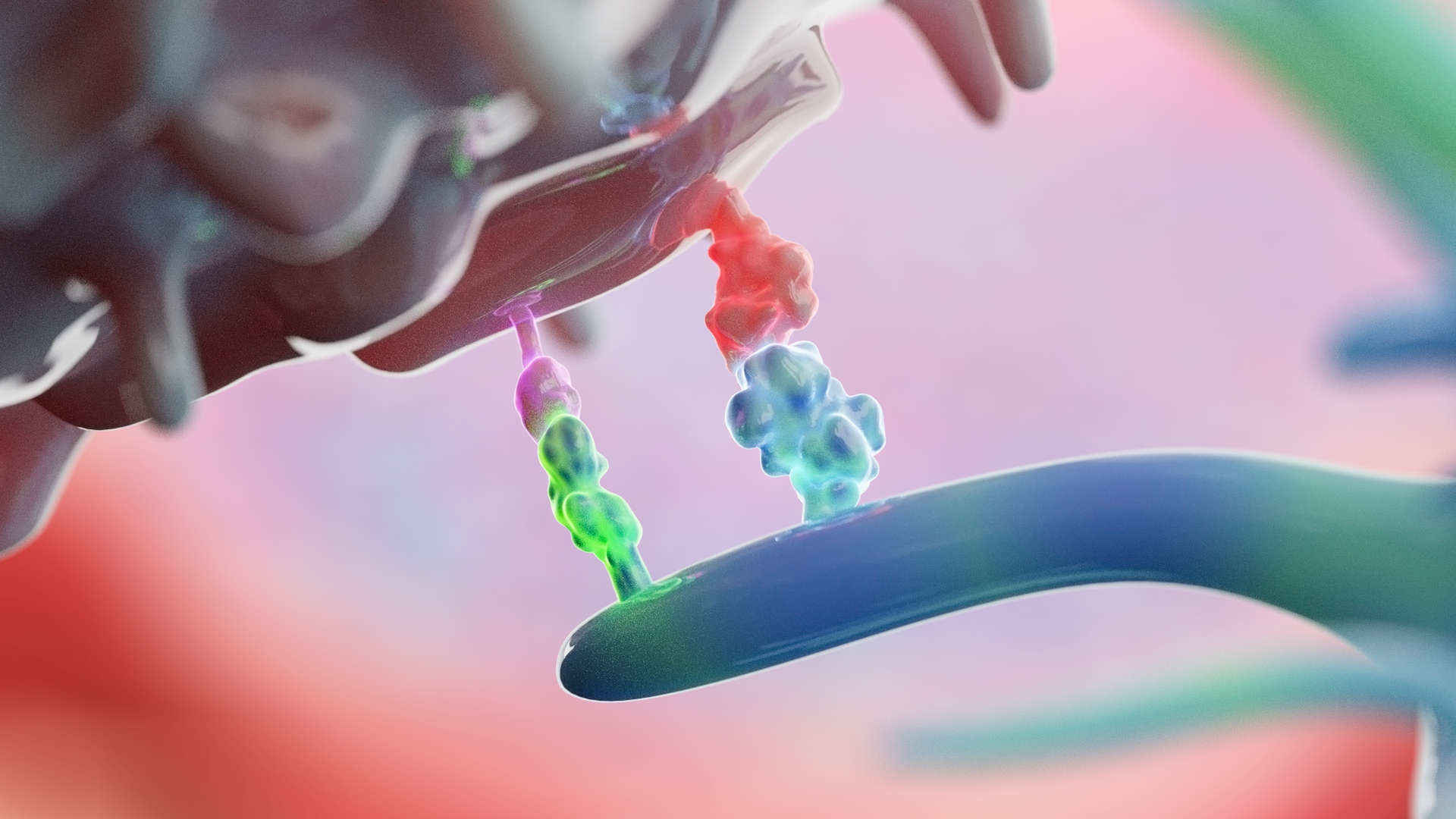 Figure 1. 3D rendering of immune checkpoint molecules on t cell and dendritic cell
The main goal of immune checkpoint detection is to study the expression patterns and functional regulation of these molecules, thereby uncovering their role in disease mechanisms. For instance, by detecting the expression of the PD-1/PD-L1 pathway, one can assess how tumor cells use immune evasion to gain survival advantages. Today, immune checkpoint inhibitors (such as anti-PD-1 and anti-CTLA-4 antibodies) have become crucial in cancer treatment, making immune checkpoint detection a key step in drug development and therapeutic strategy optimization. This has become a research hotspot in immunology in recent years.
Creative Diagnostics has been deeply involved in biological testing technology. Through independent innovation, we have developed a high-throughput detection platform based on protein array technology, widely used in immune checkpoint molecule detection. Our protein array technology, combined with various advanced detection methods, provides comprehensive and efficient solutions to enhance the depth and breadth of immune checkpoint research.
When to consider choosing our protein arrays?
1. Cancer Immunotherapy Research
Our protein array technology enables precise detection of the expression levels of PD-1/PD-L1, CTLA-4, and other molecules, providing valuable data for tumor immune microenvironment research. Additionally, we can use multi-molecule detection to reveal the synergistic effects of multiple key molecules in the immune suppression network, offering insights for immune therapy target development.
2. Multi-checkpoint Molecule Co-detection
Through a single experiment, you can simultaneously analyze multiple immune checkpoint molecules such as TIGIT, LAG-3, and TIM-3, mapping out a comprehensive immune regulation network. Ideal for who is studying the synergistic effects of multiple checkpoint molecules.
3. Precise Immune Monitoring
In the study of chronic diseases and autoimmune diseases, our protein array technology can help uncover abnormal signaling pathways in immune cell activation and regulation, driving the development of novel immune therapies.
4. Drug Development Support
Our detection platform can quickly assess the activity and efficacy of immune checkpoint inhibitors, providing strong data support for preclinical research and drug screening.
Array Example
| Panel | Example Marker Combination | Target Application | Possible Sample Preparation |
| Panel A (Core Checkpoints) | PD-1, PD-L1, PD-L2, CTLA-4 | - Evaluate immunotherapy targets (anti-PD-1, anti-CTLA-4) - Monitor therapy-induced changes in PD-L1 expression in tumors or serum | - In vitro: Culture supernatants or cell lysates from T cells, tumor cells, or DCs - In vivo: Serum/plasma (soluble checkpoints), tumor biopsies (if protein extracted), PBMC lysates or surface-shed molecules |
| Panel B (Extended Inhibitory) | LAG-3, TIM-3, TIGIT, VISTA, B7-H4, B7-H3 | - Identify T cell exhaustion patterns in cancer or chronic infection - Discover alternative checkpoint upregulation upon PD-1 blockade | - In vitro: T cell cultures from chronic infection models, supernatants - In vivo: Serum, tumor microenvironment (TME) fluid, ex vivo TILs (tumor-infiltrating lymphocytes) or lymph node cell lysates |
| Panel C (Costimulatory) | CD28, ICOS, 4-1BB, OX40, GITR, CD80, CD86 | - Assess balance between inhibitory and stimulatory pathways - Investigate combination therapies (e.g., checkpoint inhibitors + costim agents) | - In vitro: Co-culture supernatants (DC + T cells), or cell lysates for membrane-bound markers - In vivo: Serum (some costim can be shed), tissue extracts, or ex vivo activated T cells from spleen/lymph nodes |
Our Biological Protein Array Technology Leads the Way
High-throughput and Multi-indicator Detection
Our advanced platform supports multi-indicator co-detection, enabling multiple checkpoint molecules to be analyzed in a single test, significantly reducing experimental time and cost.
Ultra-high Sensitivity and Specificity
Combined with laser confocal and near-infrared fluorescence labeling technology, our protein arrays achieve picomolar-level detection sensitivity with high molecular recognition specificity, ensuring accurate and reliable results.
Fully Customizable Services
We offer flexible, customized solutions to meet different customer research needs, including array design, label optimization, and experimental process development, ensuring the best fit for basic research, clinical applications, and drug development.
Diverse Detection Platforms
The protein array technology can also be integrated with magnetic microparticle platforms to enable rapid detection of single indicators or specific molecules, complementing high-throughput analysis and suitable for various research and detection scenarios in hospitals, research centers, and companies.
Wide Applications Across Fields:
- Oncology Used to study the expression patterns of immune checkpoint molecules in the tumor microenvironment, evaluate the efficacy of immunotherapy, and develop personalized treatment strategies.
- Autoimmune Diseases Reveals immune regulatory mechanisms in diseases like rheumatoid arthritis and lupus, offering new approaches for diagnosis and treatment.
- Infectious Disease Research Analyzes immune suppression pathways in chronic viral infections (e.g., HIV, HBV), advancing breakthroughs in treatments for virus-related diseases.
- Basic Immunology Research Systematically analyzes the functional regulatory networks of immune cells such as T cells, NK cells, and monocytes, helping discover new biomarkers and therapeutic targets.
- Drug Development Supports full-process research, from early target discovery to preclinical validation, providing an efficient drug development platform for pharmaceutical companies.
…
Creative Diagnostics is committed to pushing the boundaries of life science research.
Why Choose Us?
- Outstanding Technical Support
- One-stop Solution
- Flexible Collaboration Models
We look forward to collaborating with you to provide powerful support for your research and innovation. Want to learn more about our technology/products? Please contact us to start your journey!
- Custom Protein Arrays
- Drug Screening and Toxicity Assessment
- Immunoprofiling
- Oncology Drug Screening
- Signaling Pathway Analysis
- Spatial Multi-Omics and Targeted Proteomics
- Vaccine Development
- Antibody Profiling
- Protein-Protein Interaction Studies
- Allergen Screening
- Protein-Nucleic Acid Interaction Analysis