Signaling Pathway Analysis
Inside cells sequential molecular interactions make up cell signaling pathways which produce specific products or change how cells behave. Researchers use Signaling Pathway Analysis to investigate cellular signal transduction networks to determine cell responses to external stimuli and their effects on cell behavior and function. Studying signaling pathways helps to uncover essential life principles and disease mechanisms which assists in developing new drugs.
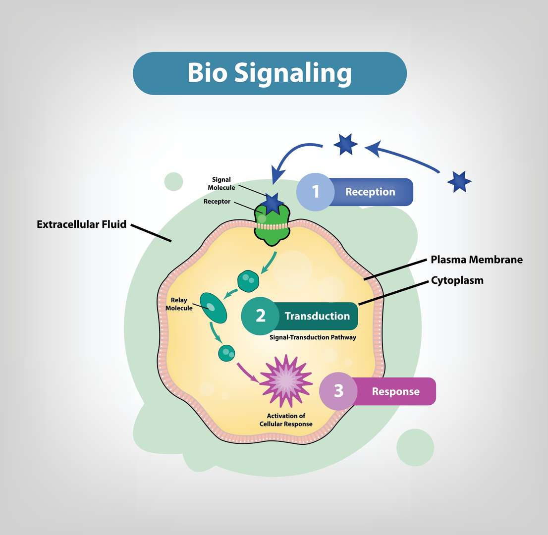
Our Signaling Pathway Analysis Services
We provide detailed signaling pathway studies using advanced technology platforms that support your research into signal transduction mechanisms.
- Phosphorylation Status Detection: The service analyzes specific protein phosphorylation levels to evaluate signal transduction pathway activation status. Learn More
- Pathway Activation/Inhibition Profiling: The examination of key proteins within pathways through expression and modification analysis helps determine whether signaling pathways are activated or inhibited. Learn More
- Glycosylation Status Detection: Protein glycosylation serves as a key modification process that controls cell signaling functions. The service enables researchers to detect protein glycosylation status which offers fresh approaches to investigating signaling pathways. Learn More
- Kinase Activity Assay: Kinases function as essential enzymes which regulate protein phosphorylation events in signaling pathways. This service offers various kinase activity detection methods that help researchers study kinase roles in signaling pathways. Learn More
- Cross-Talk Between Pathways: Complex interactions exist between different signaling pathways within cells. This service studies the cross-regulation between pathways, helping you understand the overall regulation of the signaling network. Learn More
Array Example
| Panel / Focus | Example Marker Combination | Target Application | Possible Sample Preparation |
| A. Phosphorylation Status Detection | - Phospho-specific antibodies: p-ERK (Thr202/Tyr204), p-Akt (Ser473), p-STAT3 (Tyr705), p-p38 (Thr180/Tyr182), p-IKKα/β (Ser176/180), p-S6 (Ser235/236) - Total proteins for ratio calculations (ERK, Akt, STAT3, p38, IKK, S6) | 1. Measure activation of MAPK, PI3K/Akt, JAK/STAT, NF-κB, mTOR, etc. 2. Compare basal vs. stimulated or drug-treated states 3. Map dynamic phospho-changes over time | - In vitro: Cell lysates from stimulated or drug-treated cultures; use phosphatase inhibitors - In vivo: Tissue/tumor homogenates or biopsies; snap-frozen or quickly lysed in phosphatase inhibitor buffers |
| B. Glycosylation Status Detection | - Glycan-binding arrays to detect specific sugar moieties (e.g., sialic acid, fucosylation, mannose residues) - Lectin arrays (ConA, WGA, PHA-L, etc.) to identify glycosylation patterns - Glycosyltransferase or glycosidase profiling (e.g., FUT8, MGATs) | 1. Characterize glycoprotein modifications (e.g., on receptors, adhesion molecules) 2. Study how glycosylation changes affect ligand binding or immune recognition 3. Assess glycan patterns in disease states (cancer, autoimmunity) | - In vitro: Secreted glycoproteins in supernatants, or cell membrane extracts - In vivo: Serum glycoproteins, or tissue lysates for more abundant glycoproteins. Often requires specialized labeling or detection of sugar moieties |
| C. Kinase Activity Assay | - Kinase-substrate arrays: multiple peptide substrates for different kinases - Phospho-site detection for substrate readouts (e.g., GSK-3 peptide, MAPK substrate motifs) | 1. Directly measure kinase enzymatic activity (e.g., which substrates get phosphorylated) 2. Rank compound potency in inhibiting certain kinases 3. Profile broad kinome activity in disease or drug response | - In vitro: Partially purified kinase fractions or cell lysates + array of immobilized peptide substrates; measure phosphorylation signal - In vivo: Usually specialized fractionation from tissues or immunoprecipitated kinases for functional assays |
| D. Cross-Talk Between Pathways | - Multi-pathway arrays combining MAPK (ERK, JNK, p38), PI3K/Akt, NF-κB, JAK/STAT, Wnt/β-catenin, TGF-β/Smad, Notch markers - Phospho vs. total for each node | 1. Map interactions: e.g., if inhibiting EGFR drives up NF-κB or if p-Akt and p-ERK co-activate 2. Identify alternative routes enabling cell survival or proliferation 3. Guide combination therapy or multi-target approaches | - In vitro: Cell lysates from co-stimulation or multi-signal integrative experiments - In vivo: Tissue samples from complex disease models (e.g., immuno-oncology), measure multiple pathways in parallel to see synergy or cross-regulation |
| E. Pathway Activation / Inhibition Profiling | - Pathway-specific markers (e.g., for RAS/MAPK, PI3K/mTOR, NF-κB, JAK/STAT, Wnt) - Upstream RTKs (EGFR, HER2, c-Met) and downstream effectors (p-S6, p-4E-BP1, p-IkB, p-STATs) - Inhibitory proteins or negative regulators (e.g., PTEN, SHP-1) | 1. Quantify how a drug or genetic perturbation up-/down-regulates entire pathway 2. Monitor dose-response or time-course of activation/inhibition 3. Compare multiple compounds for selectivity or synergy across pathways | - In vitro: Dose- or time-course drug treatments in cell lines, measure changes in target pathway markers - In vivo: Serum or tissue from treated animals/patients, measure changes in multiple nodes to confirm on-target or off-target effects |
Our Near-Infrared Microarray Protein Chip Technology Platform
Based on independent innovation, Creative Diagnostics has combined technologies such as... to establish a unique protein chip detection platform. This platform has significant advantages in high throughput, multi-indicator joint testing, and high-cost performance.
- Special Functional Silicone Membrane: Easy to achieve high-throughput detection, with high sensitivity, easy automation, high tolerance and broad spectrum, saving detection time, and good biocompatibility.
- Near-Infrared Fluorescence Labeling: The signal is stable, easy to clean, and easy to achieve fully automatic operation.
- Laser Confocal System: High sensitivity, large linear range of determination, simple operation, easy to achieve automation, using point-by-point, line-by-line scanning, high image resolution, and no image distortion problems.
Service Features
- High-Throughput Detection: Protein chip technology can simultaneously detect multiple indicators, improving detection efficiency.
- Multi-Indicator Joint Testing: Multiple key proteins in the signaling pathway can be jointly detected to provide more comprehensive information.
- High Cost Performance: Optimize experimental procedures and reduce detection costs.
- Customized Services: Provide flexible testing solutions according to your research needs.
- Professional Data Analysis: Provide professional data analysis reports to help your research.
Service Process
- Consultation: Communicate your research needs with our technical team.
- Plan Design: We tailor an experimental plan for you.
- Sample Preparation: You provide samples or entrust us to prepare samples.
- Testing and Analysis: We use the protein chip platform for high-throughput detection.
- Data Analysis: We provide professional data analysis reports.
With advanced technology platforms, comprehensive service items, and our professional team, Creative Diagnostics is committed to providing strong support for your signaling pathway research. Our signaling pathway analysis service selection will enable you to achieve new scientific discoveries in life sciences. We are excited to join forces with you for discovering life's mysteries together.
If you have any questions or needs, please contact us.
- Custom Protein Arrays
- Drug Screening and Toxicity Assessment
- Immunoprofiling
- Oncology Drug Screening
- Signaling Pathway Analysis
- Spatial Multi-Omics and Targeted Proteomics
- Vaccine Development
- Antibody Profiling
- Protein-Protein Interaction Studies
- Allergen Screening
- Protein-Nucleic Acid Interaction Analysis