Immune Escape Mechanism Analysis
What is Immune Escape?
Immune Escape, or immune evasion, refers to the process by which pathogens, tumor cells, and other disease-causing factors use various strategies to avoid detection and elimination by the immune system. In pathogens, this mechanism includes antigen variation, escape mutations, and suppression of innate or adaptive immune functions, ultimately preventing the host immune system from recognizing and eliminating the invader. In tumors, cancer cells achieve immune evasion by downregulating antigen presentation, activating immune checkpoint pathways (such as PD-1/PD-L1), secreting immune-suppressive factors (such as TGF-β), and remodeling the tumor microenvironment. These mechanisms allow cancer cells to proliferate and metastasize while avoiding immune system clearance.
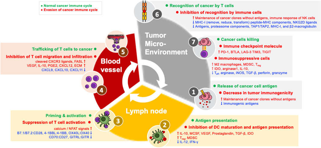 Figure 1. Immune evasion mechanisms in the cancer immune cycle. (Source: Kim SK, Cho SW., 2022)
Studying immune evasion mechanisms is crucial for understanding pathological processes, developing novel vaccines and therapeutic strategies, and optimizing existing immunotherapy research.
Creative Diagnostics use internationally leading innovative protein array platform to provide comprehensive detection from molecular to system levels for your immune evasion research, supporting disease exploration, drug development, and clinical translation.
Explore the mysteries of immune evasion starting here!
Applications of Our Protein Array in Immune Escape Mechanism Analysis
As innovators in protein array technology, we provide efficient and precise solutions for immune evasion mechanism analysis. Our protein arrays, based on a multi-parameter platform and cutting-edge technology, allow for comprehensive research on the key pathways and molecules involved in immune evasion. We assist you and your team in:
Tumor Immune Evasion Mechanism Research
Analyzing how tumor cells evade immune surveillance through downregulation of MHC expression, activation of immune checkpoints, and release of immune-suppressive factors.
Detecting molecular networks associated with immune evasion in the tumor microenvironment to support the optimization of tumor immunotherapies.
Pathogen Immune Evasion Mechanism Analysis
Detecting antigen variation, secretion of immune-suppressive factors, and host immune response regulation to provide key insights for vaccine development and infectious disease treatment.
Combination Therapy Strategy Development
Systematically evaluating the synergistic effects of immune checkpoint inhibitors and other therapies (such as antigen presentation enhancers) to provide a basis for the design of novel combination therapies.
Drug Screening and Efficacy Monitoring
Detecting immune evasion molecules to quickly assess the intervention effects of candidate drugs on relevant pathways, assisting with the full process of drug development.
Array Example:
| Panel | Example Marker Combination | Target Application | Possible Sample Preparation |
| Panel A (Checkpoint Molecules) | PD-L1, PD-1, CTLA-4, LAG-3, TIM-3, TIGIT, B7-H3, B7-H4 | - Investigate checkpoint-mediated immune suppression in tumors - Evaluate escape from immunotherapy (anti-PD-1, anti-CTLA-4) | - In vitro: Supernatants or lysates from tumor cell lines or T cell co-cultures - In vivo: Serum (soluble forms), tumor/tissue lysates from animal models or patient biopsies (if feasible) |
| Panel B (Immunosuppressive Cytokines & Enzymes) | IL-10, TGF-β, IL-35, IDO, Arg1 | - Profile immunosuppressive microenvironment - Identify cytokine-driven T cell dysfunction, Treg/MDSC expansion | - In vitro: Culture supernatants from T cells + tumor cells or DCs, after stimulation - In vivo: Serum or tumor microenvironment fluid, ex vivo restimulation supernatants |
| Panel C (Treg & MDSC Markers) | FoxP3, CD25, CD127 (Tregs), CD11b, Gr-1, CD14, CD15, S100A9 (MDSC-related) | - Quantify suppressive cell types (Tregs, MDSCs) in tumors or chronic infection sites - Evaluate mechanisms of immune evasion by these cell populations | - In vitro: Lysates from sorted Treg or MDSC cultures; supernatants if measuring secreted factors (e.g., S100A9) - In vivo: Tissue homogenates (tumor, spleen, LN), PBMC lysates, or serum for soluble markers, if present |
Technical Advantages of Our Platform
Efficient Multi-Parameter Detection
The protein array platform supports simultaneous detection of multiple immune-related targets, like immune checkpoint molecules (PD-L1, TIM-3), antigen presentation molecules (MHC-I), and immune regulatory factors (TGF-β, IL-10), significantly enhancing experimental efficiency. Its high-throughput detection capability enables the handling of large sample volumes in a single experiment, suitable for large-scale screening and frequent experimental applications.
Outstanding Sensitivity and Precision
Combining laser confocal imaging and near-infrared fluorescence labeling technology, our microarrays can precisely detect low-abundance molecules with picomolar sensitivity, while optimizing the chip surface to ensure high specificity to target molecules, significantly reducing background noise and ensuring data accuracy.
Intelligent Data Analysis and Custom Services
We provide intelligent data collection and analysis tools to help quickly extract key information from experiments. In addition, we offer flexible custom services based on client needs, including specific molecule detection, pathway analysis design, and chip content optimization, ensuring that we meet the diverse needs from basic research to industrial drug development.
Resource Efficiency and High Support
Our technology requires minimal sample volumes, allowing for multi-parameter detection with very little sample input, making it especially suitable for studies with scarce samples. The resulting data meets international standards, which can be directly used for research publication, international collaboration, or drug registration approval, helping accelerate your research into practical applications.
Why Choose Creative Diagnostics?
- Cutting-Edge Technology Assurance
- Professional Team Support
- Diverse Cooperation Models
- Efficient Delivery and Comprehensive Support
Contact Us to choose more products and services and breakthrough the possibilities of science!
Reference
- Kim SK, Cho SW. The evasion mechanisms of cancer immunity and drug intervention in the tumor microenvironment. Front Pharmacol. 2022;13:868695.
- Custom Protein Arrays
- Drug Screening and Toxicity Assessment
- Immunoprofiling
- Oncology Drug Screening
- Signaling Pathway Analysis
- Spatial Multi-Omics and Targeted Proteomics
- Vaccine Development
- Antibody Profiling
- Protein-Protein Interaction Studies
- Allergen Screening
- Protein-Nucleic Acid Interaction Analysis