Pathway Activation/Inhibition Profiling
What Is Pathway Activation/Inhibition Profiling?
Pathway Activation/Inhibition Profiling is an advanced analysis process to see if cellular signaling pathways are activated or disabled in response to either a stimulus (drugs, environmental) or another factor. It is this profiling that determines the interactions and modifications of proteins in these pathways, giving a better understanding of the dynamic mechanism that drives cell behavior. Through reverse-phase protein arrays and other protein array methods, it’s possible to quantify protein expression and post-translational modifications to enable researchers to view the full picture of pathway function.
The study of signaling pathway activation and inhibition holds significant importance because it impacts multiple aspects of biological understanding.
- Mechanisms of Disease: Misregulation of signaling systems drives numerous diseases and prominently includes cancer development. Pathway tracing allows us to detect disease progression and identify treatment targets.
- Biomarker Identification: Pathway profiling identifies biomarkers that indicate disease presence or severity to support diagnostic and prognostic processes. Activation patterns of biological pathways show correlations with different cancer types and these findings can improve targeted therapeutic approaches.
- Therapeutic Development: Once scientists have a clear understanding of which pathways respond to therapies, they can design better treatments that target those pathways directly and enhance the treatment response.
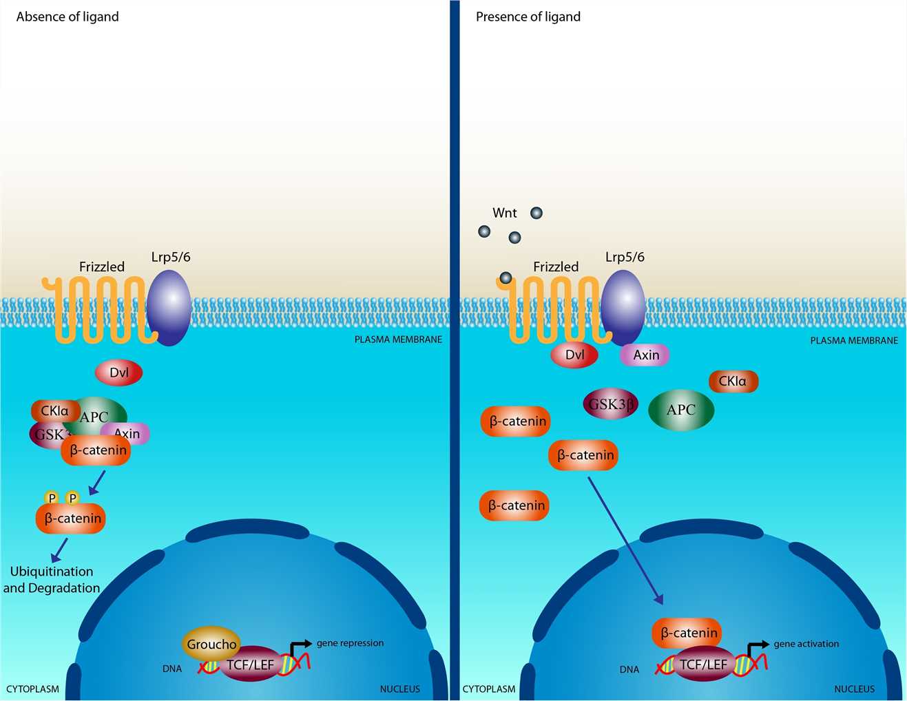
Protein Array Provides Service for Pathway Activation/Inhibition Profiling
At Protein Array, we leverage cutting-edge protein array technology to offer comprehensive Pathway Activation/Inhibition Profiling services. Our approach utilizes high-specificity antibodies to quantify proteins and their modifications across numerous samples with high sensitivity and precision. This enables us to assess:
- The activation status of key signaling pathways in response to various stimuli.
- The impact of drug candidates on specific cellular processes.
- Differences in pathway activation across different cell types or treatment conditions.
Our services are designed to support both basic research and clinical applications, facilitating a deeper understanding of cellular signaling dynamics.
The advantages of our Pathway Activation/Inhibition Profiling services include:
- High Sensitivity: The ability of our methods to detect low-abundance proteins and modifications allows them to analyze clinical samples where available material is scarce.
- High Throughput: The simultaneous processing of hundreds to thousands of samples enables us to conduct large-scale studies with statistically meaningful outcomes.
- Comprehensive Analysis: Simultaneous profiling of multiple signaling pathways allows us to construct a complete picture of cellular responses which leads to improved analysis of complex biological data.
- Robustness: Our technology has been validated across various platforms and laboratories, ensuring consistent and reproducible results.
Array Example
| Panel | Example Marker Combination | Target Application | Possible Sample Preparation |
| Panel A (Growth Factor / RTK) | EGFR (pY1068), HER2 (pY1248), AKT (pS473), mTOR (pS2448), ERK1/2 (pT202/Y204), MEK1/2 (pS217/S221) | - Evaluate whether a drug (e.g., kinase inhibitor) blocks MAPK or PI3K/Akt signaling - Check for off-target RTK activation in cancer models | - In vitro: Cell culture lysates from tumor lines ± inhibitor - In vivo: Tumor tissue homogenates (mouse/human), serum if secreted factors are relevant |
| Panel B (Inflammation / Immune) | NF-κB p65 (pS536), IκBα (pS32), STAT1 (pY701), STAT3 (pY705), STAT5 (pY694) | - Monitor cytokine-driven pathways (e.g., JAK/STAT) in immune or inflammatory conditions - Assess NF-κB inhibition or T cell polarization | - In vitro: PBMC or T cell lysates ± cytokine stimulation - In vivo: Serum, spleen or LN cell lysates, or relevant tissue (e.g., inflamed colon, synovial tissue) |
| Panel C (Stress & Apoptosis) | p38 MAPK (pT180/Y182), JNK (pT183/Y185), c-Jun (pS73), Caspase-3 (cleaved), PARP (cleaved) | - Check stress-activated MAPKs in response to toxins or UV - Determine apoptotic events in cancer therapy or immune cell death | - In vitro: Lysates from cells exposed to stress (UV, chemical) - In vivo: Tumor or tissue extracts, possibly time-course post-therapy or injury |
Protein Array's Near-Infrared Microarray Protein Chip Technology Platform
Protein Array offers a cutting-edge near-infrared microarray protein chip technology platform that enhances the capabilities of Pathway Activation/Inhibition Profiling. Our platform combines technologies such as laser confocal microscopy, near-infrared fluorescent labeling chip, and special functional silica gel matrix membrane with protein chips to improve detection sensitivity and specificity, allowing for more precise analysis of protein interactions and drug effects.
Key features of our technology include:
- Greater Sensitivity and Specificity: Near-infrared technologies provide lower detection limits than other technologies which reduce false positives and increase the reliability of screening outcomes.
- Minimal Sample Volume Required: With microarray technology, you do not need as much sample for every test, thereby making it cheaper and allows you to test harder-to-find or expensive compounds.
- Automation & High Throughput: This tool allows you to automate processes with high samples per run for more efficiency and throughput.
- Multi-Indicator Joint Detection: The platform is also applicable for multi-indicator joint detection. It also works for a variety of purposes, like enzyme inhibition experiments or protein-protein interactions, so it is really a drug finder’s all-in-one.
Application of Pathway Activation/Inhibition Profiling
Pathway Activation/Inhibition Profiling has a wide range of applications across various fields:
- Cancer Research: Understanding dysregulated pathways in cancer cells helps scientists comprehend tumor biology and create specific treatments.
- Drug Development: Analyzing drug effects on signaling pathways enables the refinement of treatment plans and the forecasting of individual patient outcomes.
- Clinical Diagnostics: Pathway activity profiling functions as a diagnostic instrument which enables clinicians to evaluate patient conditions and customize treatment options.
- Basic Biological Research: Pathway analysis of basic cellular processes leads to breakthroughs in the fields of molecular biology and biochemistry.
Leveraging the advanced near-infrared microarray protein chip technology platform from Protein Array, Pathway Activation/Inhibition Profiling services of Creative Diagnostics provide essential insights into cellular signaling networks, facilitating advancements in research and clinical applications aimed at improving health outcomes.
- Custom Protein Arrays
- Drug Screening and Toxicity Assessment
- Immunoprofiling
- Oncology Drug Screening
- Signaling Pathway Analysis
- Spatial Multi-Omics and Targeted Proteomics
- Vaccine Development
- Antibody Profiling
- Protein-Protein Interaction Studies
- Allergen Screening
- Protein-Nucleic Acid Interaction Analysis