Cell Signaling Pathway Mapping
What Is Cell Signaling Pathway Mapping?
Through cell signaling pathway mapping researchers can systematically examine networks of intracellular signal transduction pathways. We aim to discover intracellular signaling molecules and map their interactions to create detailed maps of cell signaling pathways. Through scientific research on cellular pathways we gain knowledge about how cells react to external signals and learn about their control over essential biological functions including cell growth and cell death while examining effects on tumor development.
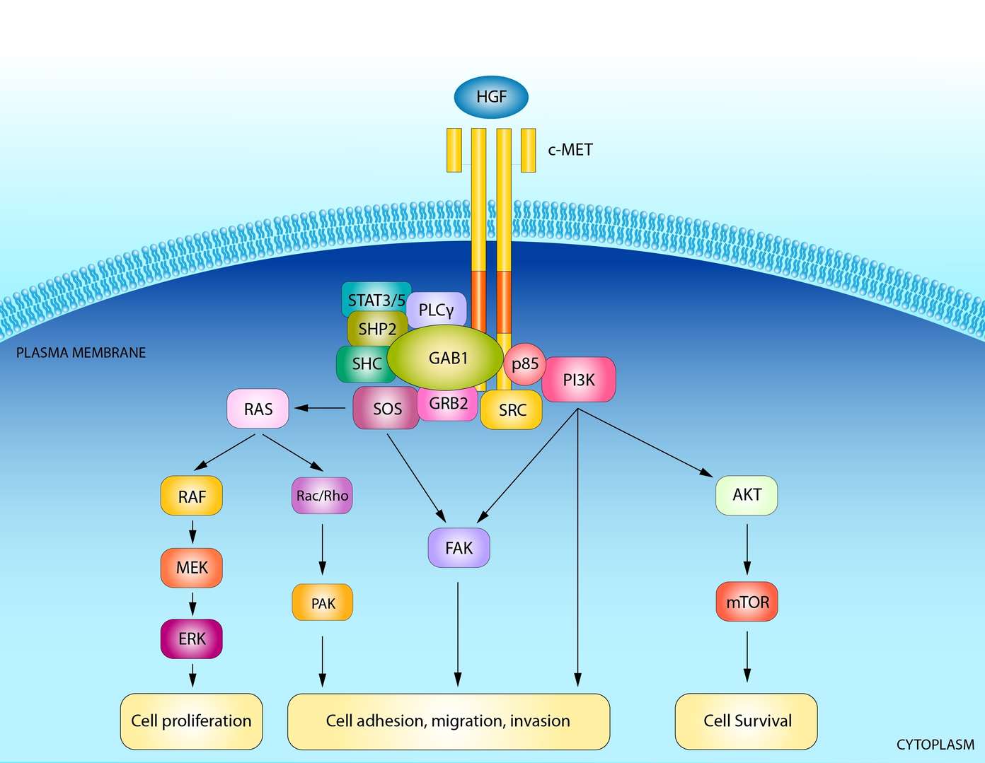
Mapping cell signaling pathways proves essential for multiple research purposes.
- Understanding Tumorigenesis Mechanisms: The observation of unusual signaling pathway behavior in cancerous cells improves our knowledge about the molecular mechanisms that govern tumor development and advancement.
- Identifying Drug Targets: Drug development strategies benefit from the discovery of specific signaling pathway nodes within tumor cells.
- Predicting Drug Efficacy: Medical experts analyze tumor cell signaling pathways to understand patient responses to specific medications.
- Personalized Treatment: Design treatment protocols individually for patients by using their unique signaling pathway maps.
- Overcoming Drug Resistance: Studying tumor cell resistance mechanisms helps create strategic approaches to defeat drug resistance.
Protein Array Services for Cell Signaling Pathway Mapping
Our Service Offerings
- Phosphorylation Analysis: Through our research on phosphorylated proteins, we can determine the routes of phosphorylation signaling.
- Kinase Activity Analysis: We can identify which signaling pathways are active by measuring essential cellular kinase activities.
- Transcription Factor Activity Analysis: We use cellular transcription factor activity measurements to figure out how signaling pathways control gene expression regulation.
- Cytokine Analysis: The levels of cytokine expression in cells serve as monitoring tools for interactions between signaling pathways and immune system mechanisms.
- Signaling Pathway Inhibitor Screening: We identify compounds designed to block specific signaling pathways as potential drug development candidates.
- Customized Signaling Pathway Analysis Solutions: Every customer receives tailored analysis of signaling pathways from our team.
- Bioinformatics Analysis: Providing professional signaling pathway data analysis and bioinformatics support to help customers deeply mine data value.
- Pathway Validation Services: Validating the reliability of signaling pathway analysis results through experiments such as Western Blot and qPCR.
Array Example
| Panel / Focus | Example Marker/Feature Combination | Target Application | Possible Sample Preparation |
| A. Phosphorylation Analysis | - Phospho-specific vs. total antibodies for MAPK (ERK, p38, JNK), PI3K/Akt (p-Akt, p-S6), JAK/STAT (p-STAT1, p-STAT3), NF-κB (p-IKK, p-IκBα) - Other key p-forms: p-PLCγ, p-SHP2, p-CREB, etc. | 1. Track dynamic activation of major signaling pathways under various stimuli (growth factors, stress, drugs) 2. Compare baseline vs. post-treatment states in disease models 3. Map multi-pathway crosstalk by measuring changes in phospho-levels simultaneously | - In vitro: Cell lysates from stimulated or drug-treated cultures, with phosphatase inhibitors in lysis buffer - In vivo: Tissue or tumor homogenates, snap-frozen or quickly lysed to preserve phospho-states |
| B. Kinase Activity Analysis | - Peptide substrate arrays: multiple consensus sequences for different kinase families (e.g., MAPKs, CDKs, Src family, etc.) - Functional readouts measuring incorporation of phosphate into substrates | 1. Directly measure kinase enzymatic activity (which substrates are phosphorylated?) 2. Rank compound potency for kinase inhibition 3. Profile broad kinome shifts under certain conditions (oncogenic transformation, drug exposure) | - In vitro: Partially purified kinase fractions or cell lysates; arrays with immobilized peptide substrates + detection of phosphate transfer - In vivo: Immunoprecipitated kinases or tissue lysates fractionated to enrich for kinase activity, then tested on arrays |
| C. Transcription Factor Activity Analysis | - Arrays with capture probes: short DNA consensus sequences for TF binding (e.g., NF-κB, AP-1, p53, STAT elements) - Antibody-based arrays to detect TF phosphorylation or nuclear translocation (p-p65, p53, c-Jun, FOXO3) | 1. Determine TF activation via DNA-binding capacity (e.g., NF-κB binding array) 2. Assess nuclear translocation and phosphorylation states that regulate TF function 3. Screen multiple TFs for changes in response to stimuli or drugs | - In vitro: Nuclear extracts from stimulated or treated cell cultures; measure TF binding or phospho TFs - In vivo: Nuclear fractions from tissues, tumors, or immune cells—requires timely sample collection and fractionation |
| D. Cytokine Analysis | - Pro-inflammatory cytokines: IL-1β, IL-6, TNF-α, IL-8 - Th subsets: IFN-γ, IL-4, IL-17, IL-10 - Chemokines: MCP-1, RANTES, CXCL10, MIP-1α - Growth factors: VEGF, FGF, PDGF | 1. Identify secreted signals influencing cell signaling loops (autocrine/paracrine) 2. Map immune context or tumor microenvironment signals 3. Correlate cytokine changes with pathway activation or drug effects | - In vitro: Culture supernatants from T cells, macrophages, cancer cells, etc. ± stimuli or drugs - In vivo: Serum, plasma, or tissue interstitial fluid from animal models or patients at multiple timepoints |
| E. Signaling Pathway Inhibitor Screening | - Multi-pathway phospho arrays (p-ERK, p-Akt, p-STAT3, p-p38, etc.) - Kinase substrate arrays to measure enzymatic inhibition - TF activation arrays if the inhibitor targets nuclear signaling (e.g., NF-κB, STAT) | 1. Screen multiple compounds for on-target inhibition of specific pathways 2. Detect compensatory activation of alternative pathways under inhibitor treatment 3. Rank or prioritize lead candidates based on potency and specificity | - In vitro: Robotic or high-throughput array systems with cell lysates or purified kinases ± drug - In vivo: Tissue or tumor samples from animals treated with candidate inhibitors, measuring phospho-levels or functional readouts |
| F. Pathway Validation | - Key nodes of interest (e.g., if a study suggests c-Met dependence, measure p-Met, downstream p-Gab1, p-ERK, p-Akt) - Final effectors in relevant pathways (p53, c-Myc, Cyclin D1 for cell cycle, FOXO for apoptosis, etc.) | 1. Confirm that a hypothesized pathway is indeed up/downregulated in a given model 2. Validate that manipulating the pathway node (via drug, gene knockdown) changes predicted downstream effectors 3. Demonstrate causality in mechanism-of-action studies | - In vitro: Cell lines with gene knockouts or overexpression, then measure array readouts - In vivo: Tumor xenografts or genetically engineered mouse models, tissue lysates for final pathway "signature" and post-drug validation |
Protein Array's Near-Infrared Microarray Protein Chip Technology Platform
We have established a unique protein array detection platform by combining laser confocal microscopy, near-infrared fluorescence labeling, and special functional silicone membrane technologies with protein chips. This platform provides strong technical support for cell signaling pathway mapping.
The platform has the following advantages:
- High Sensitivity: Able to detect low-abundance protein expression changes.
- High Accuracy: Advanced technology and strict quality control standards ensure accurate and reliable data.
- High Throughput: This technique enables simultaneous detection of multiple indicators across numerous samples.
- High Degree of Automation: Reduces human error and improves efficiency.
Applications of Cell Signaling Pathway Mapping
Multiple scientific disciplines rely on cell signaling pathway mapping as an essential tool.
- Oncology Drug Screening: Drug candidate evaluation involves studying how these compounds affect tumor cell signaling pathways to identify medications that work effectively.
- Drug Resistance Mechanism Research: Studies indicate that tumor cells build resistance to medications by altering their intracellular signaling pathways.
- Combination Therapy Strategies: Patients experience better treatment results when multiple signaling pathways receive simultaneous drug therapy intervention.
- Biomarker Discovery: The identification of biomarkers associated with certain signaling pathways aids in both tumor diagnosis and prediction of treatment outcomes.
- Personalized Treatment: Specific drugs targeting patients should be selected according to their signaling pathways' detailed maps.
Leveraging the advanced near-infrared microarray protein chip technology platform from Protein Array, Creative Diagnostics is committed to providing customers with high-quality cell signaling pathway mapping services to assist in cancer research and drug development.
- Custom Protein Arrays
- Drug Screening and Toxicity Assessment
- Immunoprofiling
- Oncology Drug Screening
- Signaling Pathway Analysis
- Spatial Multi-Omics and Targeted Proteomics
- Vaccine Development
- Antibody Profiling
- Protein-Protein Interaction Studies
- Allergen Screening
- Protein-Nucleic Acid Interaction Analysis