Biomarker Discovery
What Is Biomarker Discovery?
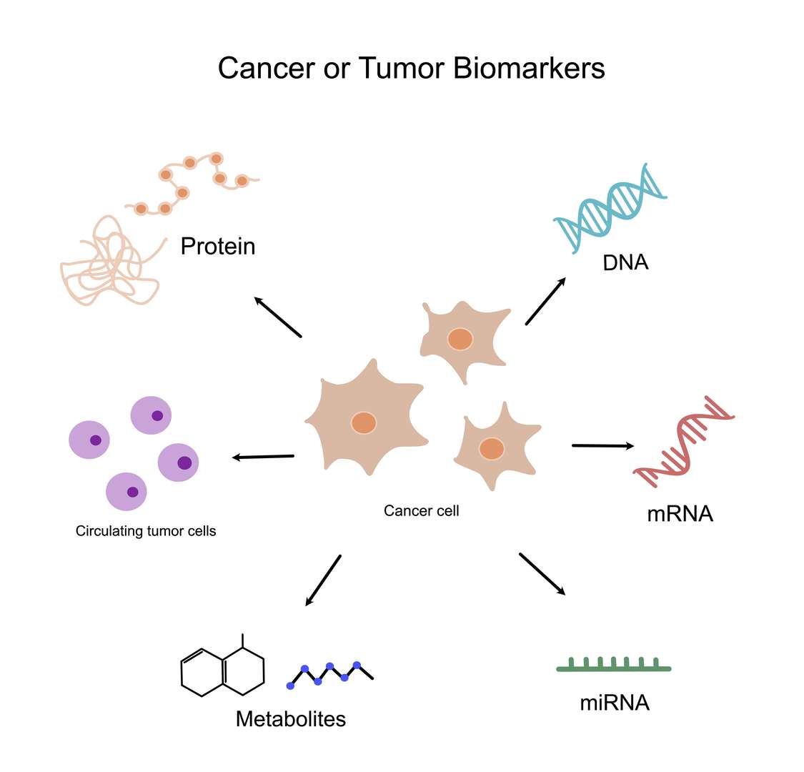
Biomarker discovery requires several steps to identify biomolecules that can function as dependable disease or physiological condition indicators. DNA, RNA, proteins, metabolites and other small molecules which demonstrate significant expression level changes throughout disease progression can serve as biomolecules for early, advanced or treatment response disease stages. Biomarkers in oncology research serve multiple purposes, from early detection of cancer before symptoms manifest through diagnosis to predicting disease progression and patient survival with prognosis tools while assessing treatment responses and developing individualized treatment approaches based on biomarker profiles. The discovery of biomarkers demands high-throughput screening operations followed by bioinformatics analysis together with stringent validation procedures to establish both reliability and clinical usefulness.
Biomarker discovery is crucial for the following aspects:
- Early Diagnosis: The discovery of biomarkers for early-stage tumors enables both early disease detection and timely intervention.
- Prognosis Prediction: The determination of tumor invasiveness and metastasis potential enables accurate patient survival predictions.
- Efficacy Evaluation: Drug treatment effectiveness evaluation needs ongoing monitoring to make necessary adjustments to treatment strategies.
- Personalized Treatment: Patient-specific biomarker profiles serve as the basis for selecting treatment approaches.
- New Drug Development: Research efforts focus on identifying novel drug targets and creating advanced treatments for cancer.
Protein Array Services for Biomarker Discovery
- Proteomics Analysis: The use of mass spectrometry techniques allows researchers to conduct thorough protein expression profile analysis which leads to biomarker discovery.
- Multi-Omics Integrated Analysis: For comprehensive cancer biological mechanism understanding scientists need to combine proteomics data with genomics data and miRNAomics data together with metabolomics data.
- Customized Biomarker Screening Solutions: Our biomarker screening solutions are designed to address the unique requirements of every customer.
- Bioinformatics Analysis: Our bioinformatics support services combined with expert data analysis enable customers to extract maximum value from their data.
- Validation Services: Our validation services employ ELISA and IHC techniques to assure biomarker reliability.
Array Example
| Panel / Focus | Example Marker/Feature Combination | Target Application | Possible Sample Preparation |
| A. Cancer Biomarker Discovery | - Cell-surface antigens: EGFR, HER2, PD-L1, MUC1, EpCAM, CD44 - Secreted markers: CEA, CA125, AFP, VEGF, IL-8 - Intracellular: p53, KRAS variants, PTEN, Rb, Cyclin D1 | 1. Identify novel or known tumor-associated proteins that correlate with cancer progression or response to therapy 2. Screen for differential expression in tumor vs. normal 3. Discover potential blood-based or exosomal biomarkers for non-invasive diagnosis | - In vitro: Cell line lysates or supernatants (tumor vs. normal lines) - In vivo: Tissue homogenates (tumor vs. adjacent normal), patient serum/plasma for secreted/exosomal proteins, or tumor biopsies |
| B. Autoimmune / Inflammatory | - Cytokines/chemokines: IL-1β, IL-6, TNF-α, MCP-1, RANTES - Autoantigens: dsDNA, Sm/RNP, SSA/Ro, SSB/La, Scl-70 - Inflammation markers: CRP, SAA, complement components (C3, C4) | 1. Pinpoint inflammatory or autoimmune signatures (e.g., rheumatoid arthritis, SLE, IBD) 2. Differentiate disease subtypes (e.g., lupus with anti-dsDNA vs. anti-Smith) 3. Track treatment response via changes in cytokine or autoantibody levels | - In vitro: Supernatants from stimulated immune cells or co-cultures (PBMCs, macrophages) - In vivo: Patient or animal serum from autoimmune/inflammatory disease models, synovial fluid (arthritis), or cerebrospinal fluid (CNS inflammation) |
| C. Infectious Disease Biomarkers | - Pathogen-specific antigens: TB antigens (ESAT-6, CFP-10), viral envelope/nucleocapsid proteins, bacterial toxins - Host response markers: acute phase proteins (CRP, SAA), interferons (IFN-α, IFN-γ), complement activation fragments | 1. Detect infection signatures (host + pathogen proteins) 2. Screen for early or latent infection biomarkers (e.g., TB, HIV, viral hepatitis) 3. Monitor therapy success (e.g., decrease in pathogen antigen or drop in host inflammatory markers) | - In vitro: Culture supernatants from infected cells or microbe lysates (for direct antigen arrays) - In vivo: Patient or animal blood, possibly tissue-specific fluids (e.g., bronchoalveolar lavage for respiratory infections) |
| D. Neurological Disease Markers | - Neurodegeneration: Tau (phospho forms), Aβ peptides, α-synuclein, TDP-43 - Neuroinflammation: GFAP, S100β, MCP-1, cytokines in CSF - Synaptic proteins: PSD-95, synaptophysin | 1. Identify potential CSF or serum biomarkers for Alzheimer's, Parkinson's, or ALS 2. Distinguish between neurodegenerative and neuroinflammatory disorders 3. Detect early vs. late-stage changes in CNS-related proteins | - In vitro: Neuronal cell line lysates, microglia supernatants - In vivo: CSF from human patients or animal models, possibly serum if crossing blood-brain barrier; brain tissue homogenates from disease vs. control |
| E. Cardiovascular / Metabolic | - Cardiac: Troponins (cTnI, cTnT), BNP/NT-proBNP, CK-MB, myosin-binding protein C - Metabolic: adipokines (leptin, adiponectin), insulin, glucagon, inflammatory cytokines (IL-6, TNF-α), glycation end products | 1. Discover or validate markers for cardiac injury (troponins), heart failure (BNP), or metabolic syndromes (adipokines) 2. Monitor disease progression or therapy response (e.g., troponin for myocardial infarction, adiponectin for metabolic improvements) | - In vitro: Cardiac cell lines, adipocyte cultures for metabolic secretions - In vivo: Serum/plasma from patients/animals with cardiac or metabolic conditions, or post-ischemia heart tissue homogenates |
| F. Pan-Omics / Exploratory | - Large untargeted arrays with hundreds to thousands of proteins (antibody-based arrays or reverse-phase arrays) - Bead-based multiplex with high coverage of cytokines, growth factors, enzymes, structural proteins | 1. Broad screening when little is known about the disease's biomarker profile 2. Generate hypotheses and lead biomarkers for further validation 3. Cluster patient subgroups by large-scale expression patterns | - In vitro: Conditioned media or lysates from disease vs. healthy cell lines - In vivo: Serum, plasma, tissue lysates from large patient cohorts or animal models; analyzing many markers simultaneously to find correlations or patterns |
Protein Array's Near-Infrared Microarray Protein Chip Technology Platform
Protein Array has established a unique protein array detection platform by combining laser confocal microscopy, near-infrared fluorescence labeling, and special functional silicone membrane technologies with protein chips. This platform provides strong technical support for biomarker discovery.
The platform has the following advantages:
- High Sensitivity: Able to detect low-abundance protein expression changes.
- High Accuracy: Advanced technology and strict quality control standards ensure accurate and reliable data.
- High Throughput: The system enables concurrent detection of numerous samples along with multiple indicators.
- High Degree of Automation: Reduces human error and improves efficiency.
Applications of Biomarker Discovery
The discovery of biomarkers serves as a key tool across multiple application areas.
- Early Tumor Diagnosis: Scientists search for biomarkers which enable early tumor detection through diagnostic procedures.
- Tumor Subtyping: The classification of tumors based on their biomarker characteristics guides clinical treatment decisions.
- Drug Target Discovery: The research aims to discover new drug targets while also developing improved anti-cancer treatments.
- Companion Diagnostics: Identifying patient populations that match specific drug treatments using screening methods.
Leveraging the advanced near-infrared microarray protein chip technology platform from Protein Array, Creative Diagnostics is committed to providing customers with high-quality biomarker discovery services to assist in cancer research and drug development.
- Custom Protein Arrays
- Drug Screening and Toxicity Assessment
- Immunoprofiling
- Oncology Drug Screening
- Signaling Pathway Analysis
- Spatial Multi-Omics and Targeted Proteomics
- Vaccine Development
- Antibody Profiling
- Protein-Protein Interaction Studies
- Allergen Screening
- Protein-Nucleic Acid Interaction Analysis