Membranous nephropathy (MN) was first described in 1959. In 1959, Heymann et al. first established the classic MN rat model-Heymann nephritis model. And then, they found that the pathogenesis of MN is the deposition of immune complexes in the podocytes in situ, and the target antigen is megalin(the rat podocyte antigen). However, megalin does not exist in epithelial cells of patients with MN, and no anti-megalin antibodies are detected in the serum. Until 2002, the first podocyte antigen of human MN, neonatal neutral endopeptidase (NEP), was first discovered in neonates with membranous nephropathy. The neutral endopeptidase of maternal antibodies crosses the placenta and binds to fetal podocytes, and then forms an immune complex in the epithelium, causing diseases.

Later, the researchers proved that M-type phospholipase A2 receptor 1 (PLA2R1) is the main human target antigen of IMN. Anti-PLA2R1 antibodies are found in 70% of IMN patients. In 2014, scientists discovered THSD7A antibodies in the sera of patients with PLA2R1-negative IMN. The discovery of PLA2R1 and THSD7A autoantibodies has made scientists more convinced that the immune system plays an important role in the pathogenesis of MN. At present, MN is considered to be an autoimmune disease, mainly caused by autoantibodies that recognize the target antigen of glomerular podocytes. The dense electronic deposits and epithelium under the foot processes can activate the complement system to form membrane attack complexes and cause podocyte damage. Systematic detection of target antigens is the key to revealing the pathogenesis of MN. The discovery and research of potential target antigens in the pathogenesis of MN may help the specific and targeted diagnosis and treatment of MN patients.
PLA2R Gene
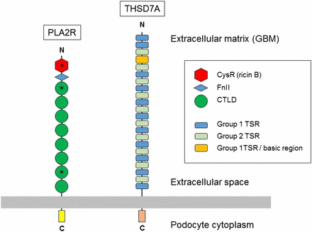
M-type phospholipase A2 receptor 1 (PLA2R1) is a type I transmembrane receptor that belongs to the mammalian mannose receptor family and is mainly expressed in podocytes. In situ immune complexes are formed by PLA2R autoantibodies expressed by IMN patients bound to the surface of podocytes. Subsequently, the complement system will be activated through the bypass and mannose-binding lectin pathway, resulting in the formation of C5b9 attack membrane complexes, thereby damaging the podocytes.
Clinical studies have found that serum samples from 26 (70%) of 37 patients with idiopathic but not secondary membranous nephropathy specifically identified the presence of non-reduced glomerular extracts 185-kD glycoprotein. Mass spectrometry of the reactive protein band identified the M-type phospholipase A2 receptor. The reactive serum specimen recognizes recombinant PLA2R and binds to the same 185-kD glomerulin as the monospecific antibody. Anti-PLA2R autoantibodies in serum samples of patients with membranous nephropathy are mainly IgG4, which is the main immunoglobulin subclass in glomerular deposits. Further research found that PLA2R is normally expressed in human glomerular podocytes and co-localizes with IgG4 in the glomerular immune deposits of patients with membranous nephropathy. Studies have found that IgG can be eluted from these deposits in patients with idiopathic membranous nephropathy, but not membranous lupus or IgA nephropathy. The results of the study indicate that most patients with idiopathic membranous nephropathy have antibodies against conformation-dependent epitopes in PLA2R.
THSD7A Gene
Thrombospondin type 1 domain 7A (THSD7A) is expressed in placental vascular endothelial cells and plays a role in endothelial cell migration and angiogenesis. A previous study evaluated the expression of THSD7A by immunofluorescence staining of kidney biopsy samples and concluded that THSD7A is expressed in podocyte foot processes.
The THSD7A antibody was detected by Tomas et al. In 2014, anti-PLA2R1 was negative in the serum of European and Boston IMN patients. At the same time, immunohistochemistry of kidney biopsy samples showed that THSD7A is a 250 kDa glomerulin that is localized in podocytes. Studies have shown that THSD7A is the second autoantigen involved in the pathogenesis of adult IMN. In this study, 15 of the 154 patients with idiopathic membranous nephropathy without anti-PLA2R1 antibodies had serum samples that responded to THSD7A. IgG4 is the main anti-THSD7A autoantibody identified in these patients, although other subtypes are weakly present in most serum samples. Immunofluorescence staining of kidney biopsy samples from healthy controls showed linear glomerular expression of THSD7A and co-localized with podocyte foot processes, but not with glomerular basement membrane or endothelial cells.
