
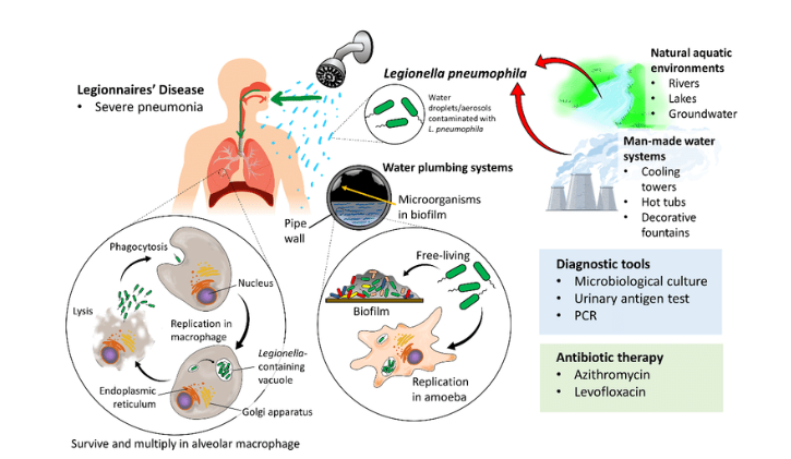
Legionnaires’ disease (LD) first broke out on a large scale at a military rally in Philadelphia in 1976, killing 34 people. Since then, there have been outbreaks or sporadic transmission of Legionnaires’ disease in the community, in hospitals, and in travel. Legionella is a facultative intracellular gram-negative bacillus that widely exists in water, soil, artificial water systems and other environments, and is mostly found in artificial water environments. Legionella bacteria mainly exist in cold water, but in recent years, Legionella bacteria have been frequently isolated from hot water systems. Legionella pneumophila (LP) is the main Legionella species that causes Legionnaires’ disease. Monitoring Legionella in the environment and effectively detecting its pathogenic ability is of great significance to protect public health and reduce losses.
Molecular Biology Methods
There are multiple virulence genes in Legionella pneumophila, including mip, ira, lvr, ot, icm, lvh, cpx, and rtxA. Mip-encoded protein can resist intracellular killing against mammalian and protozoan phagocytes; cpxRA regulates the essential factor for the secretion of effector proteins by the type IVB secretion system; the methyltransferase encoded by the iraA locus is required for bacterial intracellular infection The hypothetical iron-peptide transporter encoded by the iraB locus can be involved in the uptake of high-valent iron by means of iron-loaded peptides; the lvr gene is responsible for the regulation of lvh family secreted proteins; rtxA encodes proteins related to adhesion, cytotoxicity and pore formation. Lvh encodes a protein derived from the type IVA secretion system, which helps to bind virulence; dot/icm encodes a protein derived from the type IVB secretion system, which is responsible for the replication of Legionella in host cells. Virulence genes are distinct regions of DNA that are present in the genome of pathogenic bacteria but not present in the same or related non-pathogenic strains. The pathogenicity of Legionella is closely related to its virulence, and its pathogenicity is determined according to the amplification results of pathogenic virulence genes. The polymerase chain reaction (PCR) is a molecular biology technique used to amplify the target nucleotide sequence, and the target gene to be amplified can be amplified several million times within 2 to 3 hours. After the reaction, the amplified product was separated by agarose gel electrophoresis, and the result was positive, that is, the existence of the gene was confirmed. Although the quality of template nucleic acid extraction is not high and false negative results may occur, this method has strong specificity and high sensitivity, and is the basis for other molecular biology experiments.
In addition, PCR-based pathogenicity detection methods for Legionella include multi-locus variable number tandem repeat analysis to detect virulence genotype, micro-DNA microarray to detect Legionella virulence genes, and use of genetic markers to identify Legionella clinical and strains environmental conditions.
Immunology-based Assays
ELISA
Enzyme-linked immunosorbent assay (ELISA) is a simple and rapid technique for the detection and quantification of antibodies or antigens attached to solid surfaces. The substrate to which the enzyme is added produces a color change or light signal that correlates to the amount of antigen present in the original sample. Therefore, the total amount of antigen in the sample can calculate based on the direct relationship between the intensity of the color or the chemical signal and the amount of antigen in the sample amount or concentration. This method is simple to operate and has high sensitivity. However, the method has high requirements for technicians to operate. In addition, recombinant antigens are used in the method, and the specificity is not high and false positive results are likely to occur. A key feature of the pathogenesis of L. pneumophila is the rapid influx of neutrophils into the lung, and IL-1α is a key initiator of neutrophil recruitment to the lungs of L. pneumophila-infected mice. The researchers used this technique to detect the amount of IL-1α released from mice infected with Legionella pneumophila and found that neutrophil recruitment was in response to the virulent Legionella pneumophila, and determined the relationship between the amount of IL-1α released and the strain.
Western blot
Western blotting is a method based on the specificity of antigen-antibody binding proteins. The mixed antigen samples are separated by one-dimensional or two-dimensional electrophoresis on the gel plate, and the antigen components in the gel are transferred to the blotting paper under the natural adsorption force of the blotting paper or other external forces, and then the immobilized matrix membrane is combined with the blotting paper. And then, antigen-immobilized matrix membranes were detected and analyzed by substrate development and autoradiography. Western blot analysis is simple, time-saving and labor-saving, and can identify multiple proteins at the same time. Previous studies have found that flagella and their secreted proteins contribute to the movement, host finding, biofilm colonization and virulence of L. pneumophila. Bacterial virulence can thus be determined by detecting the L. pneumophila flagella and their secreted proteins.
LDH Cytotoxicity Assay
Lactate dehydrogenase (LDH) is a stable protein that exists in the cytoplasm of normal cells. When the cell membrane is damaged, LDH is released outside the cell. There is a good correlation between the cytotoxicity of LDH and the blood concentration of animal mortality and human death. The LDH cytotoxicity detection kit can be used to detect the activity of LDH in the cell supernatant on a 500nm microplate reader to determine the degree of cell damage, thereby realizing the detection of the toxicity of Legionella. This assay is sensitive, simple to operate, and accurate; however, the assessment of cytotoxicity is based on quantification of cell damage, so it is necessary to determine the optimal amount of cells.