
Porcine pseudorabies (PR) is an acute infectious disease of pigs caused by porcine pseudorabies virus (PRV). PRV belongs to the herpesviridae family, the alphaherpesviridae subfamily, and the genus porcine herpesvirus, which is a linear DNA virus. PRV can cause vomiting, high fever and neurological symptoms in piglets, high mortality, reproductive failure in sows, and latent infection in adult pigs after high fever symptoms are tolerated. Because PR spreads quickly and is harmful, OIE lists it as a disease that must be reported. PRV has the characteristics of latent infection. After the virus infects the host through the oral, nasal and respiratory tract, it can be latent in the peripheral nervous system, and no new virus particles are produced at this time. When changes in external conditions cause animal stress, PRV will be activated to cause the host to excrete toxins, infect pigs and even cause PR outbreaks. The feature of latent infection is the main reason why PRV is difficult to eliminate.
Virus Structure
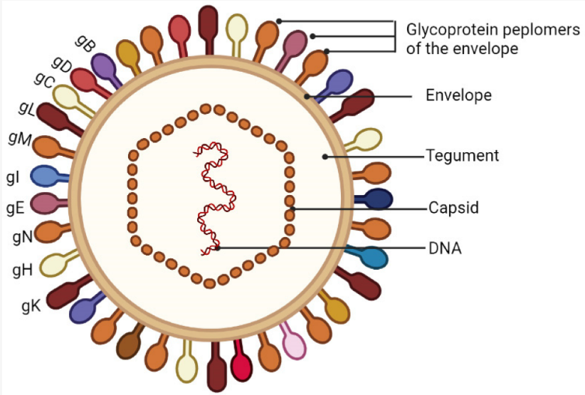
PRV virus is round or oval in shape, and is composed of double-stranded DNA, 20-hedral nucleocapsid, envelope, and lipid bilayer membrane from inside to outside. The icosahedral nucleocapsid consists of 162 capsomers, including 150 hexons and 12 pentons, with a capsid diameter of about 110nm-150nm. The envelope is located between the capsule and the capsid and is composed of a double layer of proteins, and the inner and outer component proteins are different. The envelope is located in the outermost layer of the virion and consists of 11 glycoproteins and 4 non-glycoproteins. The envelope plays an important role in the process of PRV infection of host cells. The diameter of the mature virus particles in the cytoplasm is 150-180 nm, and there are radially arranged fibers about 8-10 nm long on the surface of the capsule.
Gene Structure
The PRV genome is a linear double-stranded DNA with a size of about 150 kb and a G+C content of 74%. It has a long unique region (UL) of 110 kb and a short unique region (US) of 10 kb. Both ends of the US region are inserted with ends of 15 kb repeated Sequences (TRS) and Internal Repeated Sequences (IRS). Because the UL and US regions can be oriented in the same or opposite directions, there are two isomers of PRV, and both isomers are infectious. The origin of replication of the PRV genome, one located in the UL region is called OriL, and the other is called OriS, which is repeated in the IRS region; the PRV genome contains 73 genes and 67 open reading frames, which can encode 70 to 100 kinds of proteins. Including capsid protein, envelope glycoprotein and various enzymes, but half of them are non-essential proteins for PRV replication, so PRV can be used as the carrier of foreign genes.
Major Genes and Their Functions
TK Gene
The TK gene is located in the UL23 region, with a full length of 963 bp, encoding a total of 321 amino acids, and is a non-essential gene for PRV replication. The TK gene is one of the PRV virulence genes, and the thymidine kinase encoded by the TK gene is required for the replication of PRV in resting cells. The latent infection ability of the virus lacking TK gene is weakened in neural tissue, but it does not affect the immunogenicity of the virus. Therefore, the TK gene can be used as a target gene for gene deletion vaccines.
gE Gene
The gE gene is located in the US region. The full-length gene is 1740 bp and encodes 577 amino acids. It is a non-essential gene for PRV replication. The gE gene is one of the virulence genes of PRV. The gE protein can promote the fusion of PRV with cells and mediate the spread of the virus between cells. PRV lacking the gE gene can only infect the first-order trigeminal nerve and the sympathetic nerve that regulates the nasal mucosa. It cannot spread infection of second-order ganglia and sympathetic neurons.
gB Gene
The gB gene is located in the UL region, the full length of the gene is 2800 bp, encoding 913 amino acids, and it is an essential gene for PRV replication. The gB protein is involved in the fusion of the virus with the cell membrane and can also induce humoral immune responses. Similar to the three types of fusion proteins, gB protein has a trimeric fold, a dimer fusion ring, and an α-helix structure. The gB protein binds to the cell membrane in a cholesterol-dependent manner through the fusion ring.
gI Gene
The gI gene is located in the US region, the full length of the gene is 1053 bp, encoding 351 amino acids, and it is a non-essential gene for PRV replication. The gl protein is an envelope protein that facilitates the spread of viruses between cells. gI and gE usually exist in the form of a non-covalent bond complex, and the gI protein can promote the secretion of gE protein in the endoplasmic reticulum and ensure the correct glycosylation of gE protein.
gD Gene
The gD gene is located in the US region. The full-length gene is 1300 bp and encodes 402 amino acids. It is a non-essential gene for PRV replication. The gD protein is an envelope protein, which is highly conserved in PRV and has good immunogenicity. The gD gene is necessary for PRV to attach to cells. PRV lacking the gD gene cannot infect cells.
IE180 Gene
The IE180 gene is located in the IRS and TRS regions, respectively, encoding 1460 amino acids with a protein size of about 153 ku. The IE180 gene is the only immediate early gene in PRV that can be independently transcribed. The accumulation of IE180 protein can initiate the transcription of other genes of the virus. The PRV IE180 protein has a high degree of similarity with some regions of the herpes simplex virus type I (HSV-1) ICP4 protein, and the two are partially functionally complementary.
EP0 Gene
The EP0 gene is located in the UL region and can encode 1230 amino acids with a protein molecular weight of about 45 ku. It is a non-essential gene for PRV replication. EP0 gene has a transactivation effect on the early gene IE180, which can promote the expression of IE180 protein. EP0 protein can act together with IE180 protein to activate the transcription of TK gene and gG gene. Protein, importin α1, α3, and β1 are jointly regulated. The replication ability of PRV lacking the EP0 gene is reduced in cells, but it does not affect PRV virulence.
UL41 Gene
The UL41 gene is an essential gene for PRV replication. The UL41 gene encodes the host shutdown protein (vhs) with a protein molecular weight of about 40 ku. The vhs homology between PRV and HSV-1 is 38.4%. The vhs protein has ribonuclease activity both in vitro and in vivo, which can degrade the mRNA of host cells and inhibit the expression of host genes. Vhs can cleave the translation initiation factors eIF4H and eIF4B in the downstream region of IRES and enhance its ribonuclease activity. However, in PRV, vhs protein is not the only viral protein that can inhibit host gene expression. PRV lacking UL41 gene still has a significant inhibitory effect on host gene expression. At the same time, deletion of UL41 gene will also significantly reduce the replication ability and infectivity of PRV in vitro and in vivo.