
Hepatocellular cancer (HCC) is a clinically common malignant tumor of the digestive tract, which is seriously harmful to human health, and according to the survey, liver cancer accounts for the fifth largest incidence of tumors worldwide and the third mortality rate. At present, liver cancer in the early stages has no diagnostic methods, when examined for late stages of hepatic cancer, the majority of patients use surgical treatment, the surgery removal effect is poor, postoperative recurrence rate, transfer rate is high, to the patient’s prognosis and quality of life has a serious impact. Early diagnosis of liver cancer is important for the prognosis and survival of patients. HCC screening is currently clinically predominantly done with methylprotein (AFP), but long-term clinical practice has shown that it has a sensitivity of only 39% to 64%, and a specificity of 76% to 91% as an early diagnosis of liver cancer. Therefore, a new serum tumor marker is needed for early diagnosis of HCC patients. Serum abnormal blood clotting is a very high sensitivity and specificity in liver cancer diagnosis diagnostic indicator first by Liebman and others, which mainly refers to vitamin K deficiency or antioxidant II induced protein (PIVKA-II) that makes it difficult for liver cells to synthesize coagulation factors.
Biological Functions of PIVKA-II
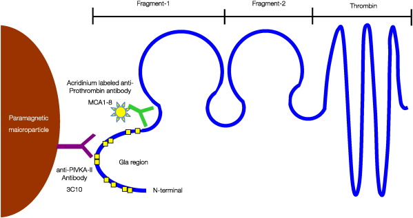
Figure 1. Assay principal of the ARCHITECT PIVKA-II.
PIVKA-II is an abnormal blood clotting enzyme, but its non-coagulation activity, which is mainly synthesized by the liver, is associated with liver cell disease. Patients with liver cancer caused an abnormality in liver cells to lack a coagulation precursor, increasing the production of PIVKA-II.A large number of clinical studies have confirmed that PIVKA-II can be used as a screening and prognostic indicator for liver cancer. However, the mechanism of overexpression of PIVKA-II in liver cancer tissue, and why tumor proliferation is associated with PIVK-II, is not fully understood, and existing basic research findings help researchers to better understand the role PIVKI-II plays in the occurrence and development of liver cancers.
PIVKA-II Reproductive Effects on Liver Cancer Cells
The structure of PIVKA-II contains two ring-shaped structural domains that are similar to the growth factor of liver cells. In hepatic cell growth factor (HCF), the ring-shaped structural domain contributes to the stable binding of HGF with the receptor (Met), thereby inducing cell proliferation. The mechanism for PIVKA-II to promote cell proliferation, especially in liver cancer cells, may be that it binds with Met to activate downstream pathways, thereby inducing prolifération. The Suzuki et al study found that the levels of DNA synthesis increased significantly in the liver cancer cell line with the addition of purified PIVKA-II. This phenomenon is more pronounced in cell lines that do not produce PIVKA-II, all of which suggest that PIVKA-II can induce the growth of liver cancer cells. Further studies have shown that after PIVKA-II is combined with Met, it activates the Met-Janus laser signal transmitting and transcribing the activation factor-3 pathway, inducing HCC cell proliferation 4.The above results suggest that the mechanism of action of PIVKA-II may be similar to that of HGF. The researchers analyzed the effect of PIVKA-II on liver cancer cell proliferation by changing the amount of secretion of PIVKA-II, injecting vitamin K2, significantly reduced the expression of PIVA-II in hepatic cancer cell line, thereby evaluating the reduction of PIVKA-II to changes in liver cancers cell line. Experimental data showed that a decrease in PIVKA-II expression reduced the degree of malignancy of the liver cancer cell line, and vitamin K2 inhibited the growth of the HCC cell line in a dose-dependent manner. Analysis found that the intramuscular injection of vitamin K2 in naked rats significantly reduced the weight after transplantation of the tumor isotope and the serum PIVKA-II water level, while no apparent acute or delayed toxic effects were observed. The findings show that controlling the levels of PIVKA-II may inhibit cancer progression, demonstrating a link between PIVKA-II and the biological behavior of the tumor.
Angiogenesis Effect of PIVKA-II on Liver Cancer Cells
PIVKA-II can enhance the vascular function around liver cancer tissue, which is another biological effect in liver cancers. Fujikawa used human umbilical vein endothelial cells(HUVEC) to analyze PIVKA-II’s vascular function, showing that PIVKI-II promotes DNA synthesis and HUVEK migration, whereas normal coagulation enzymes did not have such effects. Further studies have shown that PIVKA-II is Combined through the enzyme plug-in domain receptor (KDR), it activates the KDR-PLC-MAPK signal pathway, thereby promoting DNA synthesis and cell migration. Other studies have also obtained similar results. PIVKA-II induces overexpression of ECFR and VEGF in HUVEC cells. The above results suggest that PIVKA-II secreted by liver cancer tissue can induce vascular production of the tissue around the liver, thereby affecting the patient’s prognosis. Studies have found that PIVKA-II can promote the expression of various vascular-generating factors in liver cancer cells, including VEGF TGF-β and hFGF. Vascular formation is an indispensable phenomenon in the process of tumor occurrence and development, while closely linked to poor tumor behavior, high microvascular density can lead to recurrence of liver cancer. Therefore, PIVKA-II plays an important role in tumor processes by inducing vascular generation, and these studies all help to understand how PIVKA-II contributes to the progression of liver cancer.