
SEB poisoning can cause a series of toxic reactions and even toxic shock syndrome, with clinical manifestations of high fever, hypotension, rash, weight loss, multiple organ failure and even death, with a fatality rate of up to 50%. Sensitive and rapid detection methods are of great significance for the clinical diagnosis and prevention and control of food poisoning. At present, SEB detection methods are mainly biological methods, immunological methods, gene probe methods, instrumental analysis methods and biosensing methods.
Biological Detection Method
The detection method of Staphylococcus aureus enterotoxin B initially used relevant detection methods such as animal experiments. By intraperitoneal injection and feeding of samples with enterotoxin B added to the test animals, the abnormal physiological responses and activities of the animals were observed and recorded. The existence and toxicity of enterotoxin B were qualitatively analyzed. Such experiments are intuitive in the interpretation of the results, but the shortcomings such as poor specificity limit the application of this method.
Immunoassays Method
Immunoassays are mainly based on the principle of specific binding of antigens and antibodies, including immunoagglutination assays, agarose diffusion assays and enzyme-linked immunosorbent assays. However, the detection sensitivity of these methods is only 0.1 ng/mL, and the sample preparation and detection takes a long time. In addition, this method needs to ensure that the monoclonal antibodies have a uniform structure and avoid cross-reaction with each other.
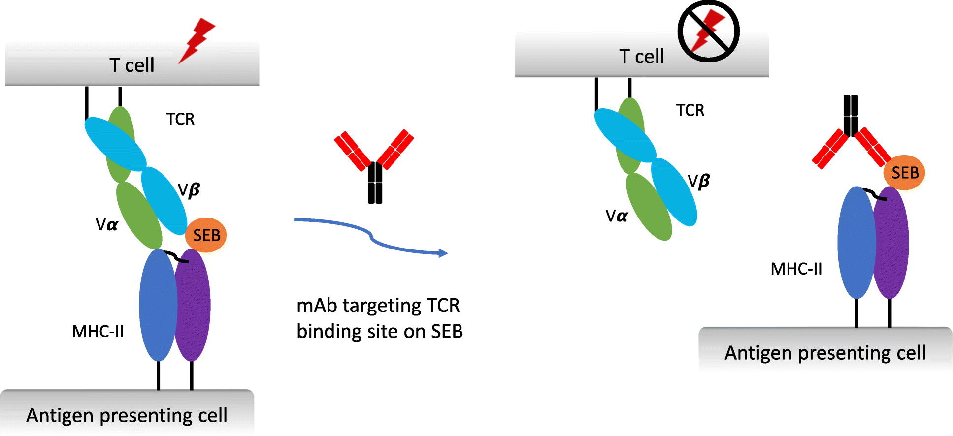
Figure 1. Potent Neutralization of Staphylococcal Enterotoxin B In Vivo by Antibodies that Block Binding to the T-Cell Receptor.
Immunoagglutination Assay
The immunoagglutination test is the agglutination produced by the combination of antigen and antibody in the presence of electrolytes. These include the reverse indirect hemagglutination test and the reverse passive latex agglutination test. The former uses the antiserum of enterotoxin B to adsorb on the surface of red blood cells of animals. When the test specimen contains SEB, hemagglutination occurs. This method can interpret the results in about 2 hours, and the detection sensitivity is greatly improved compared with the previous method; the latter adsorbs the anti-SEB specific antibody on the latex particles. When the electrolyte and SEB exist at the same time, the latex particles will agglutinate phenomenon, the detection time is about 18 h.
Immunolabeling Technology
Immunolabeling technology can be divided into enzyme-linked immunosorbent assay, radioimmunoassay and immunofluorescence detection technology. It is based on the traditional antigen-antibody reaction, by modifying fluorescent groups on the antibody, such as hydroxyfluorescein, fluoresceine isothiocyanate (FITC), water-soluble indocyanine fluorescein (cyanine-5), etc., radioisotopes, horseradish peroxidase (HRP) or alkaline phosphatase. Relevant instruments and equipment are used to output the signal intensity to achieve the purpose of detection.
- Enzyme-linked immunosorbent assay (ELISA) is an antigen-antibody reaction based on a solid-phase carrier (such as polystyrene, etc.), which catalyzes the color development of the substrate by HRP or AP, thereby amplifying the detection signal. Double-antibody sandwich and indirect competition methods are usually used to detect SEB, in which the former can obtain lower detection sensitivity and wider linear range. At present, ELISA technology has been widely used in practical detection, and targeted detection kits have been developed.
- Radioimmunoassay is based on the competitive reaction of labeled antigens (labeled with radioisotopes) and unlabeled antigens to antibodies, and is used to detect the antigen concentration of the sample to be tested. This method has high sensitivity and specificity, but it is not suitable for widespread use due to the contamination of radioisotopes.
- Immunofluorescence detection technology uses fluorescent substances (FITC, Cy5, quantum dots, up-conversion particles, etc.) as signal molecules labeled on the antibody, and indirectly determines the concentration of SEB through the fluorescence intensity generated by the labeled antibody bound to SEB, usually quantitatively determined using a fluorescence spectrophotometer.
Colloidal Gold Test Strip Detection Technology
Colloidal gold-labeled immunoassay strips mainly utilize the excellent physical and chemical properties of colloidal gold. Due to the negative charge on the surface of colloidal gold, the antibody can be immobilized on the surface by physical adsorption under alkaline conditions, and the biological activity of the antibody itself will not be affected. This method can quickly interpret the results under the naked eye, and is generally used for initial screening in the field. Among them, researchers designed a lateral flow assay (LFA) to measure SEB based on a double-antibody sandwich format on a porous nitrocellulose membrane. When a sample containing SEB is applied to the LFA device, the SEB initially reacts with polyclonal antibody-coated colloidal gold particles. These reactions produce red lines in the detection zone, the intensity of which is proportional to the SEB concentration, which can be achieved within 5 min using this method. SEB was detected at 1 ng/mL with high reproducibility.
Gene Probe Method
Enterotoxin B is transcribed and translated from the toxin expression gene sequence SEB of Staphylococcus aureus. By detecting the enterotoxin B gene of Staphylococcus aureus in food, it can be used for the positive strain of Staphylococcus aureus. Specifically, this method mainly adopts nucleic acid amplification technology, which includes polymerase chain amplification and nucleic acid isothermal amplification technology. The polymerase chain reaction (PCR) technique is mainly to design a pair of upstream and downstream primers for the target gene sequence. After adding the total DNA of the object to be detected, PCR or real-time fluorescent PCR is used to amplify the sequence of the SEB target gene, which can be quantitatively monitored by electrophoretic bands or fluorescence intensity.
Instrumental Analysis Methods
With the continuous improvement of the current detection instruments and equipment, many scholars are no longer limited to traditional immunology and other related methods, but instead use modern large-scale precision instruments for qualitative and quantitative detection of enterotoxin B. Some instrumental analysis methods can directly reveal the molecular structure of toxins, such as liquid chromatography-diode array detector, electrospray ionization, matrix-assisted laser desorption ionization, and time-of-flight mass spectrometry.
Biosensing Detection Method
Biosensor connects biochemical reactions with transducers through physical and chemical instruments, and displays them in the form of electricity. It mainly uses biological components (such as enzymes, antibodies, nucleic acids, lectins), the organic combination of biological response signal amplification (cell or tissue) and biological response signal amplification has the advantages of less sample usage and fast analysis speed, and can be used for the simultaneous detection of multiple substances. At present, the biosensors used for detection mainly include electrochemical sensors, surface plasmon resonance and piezoelectric crystal sensors.