
Inquiry Basket
 Loading ......
Human metapneumovirus (MPV) is a prevalent respiratory pathogen worldwide, causing respiratory illness similar to respiratory syncytial virus (RSV). HMPV can cause symptoms ranging from mild upper respiratory tract infections to severe bronchopneumonia. Although the virus was discovered in 2001, antibodies to MPV were found in archived human sera, indicating its circulation since the 1950s. In recent years, researchers have made significant progress in understanding MPV. They have characterized its epidemiology, developed rapid diagnostic tests, and are studying host immune responses to aid in the development of vaccines. These advancements have shed light on the impact and characteristics of MPV, facilitating better diagnosis, prevention, and management of MPV-related respiratory illnesses.
MPV, a member of the Paramyxovirus family, is an enveloped RNA virus with a single-stranded, negative-sense genome. It shares its closest relation with avian metapneumovirus type C and belongs to the Metapneumovirus genus. Within the Pneumovirinae subfamily, it is grouped together with RSV. The genome of hMPV is approximately 13 kilobases (Kb) in length and encodes nine structural proteins: N (nucleoprotein), P (phosphoprotein), M (matrix protein), F (fusion protein), M2-1 (transcription elongation factor), M2-2 (RNA synthesis regulatory factor), SH (small hydrophobic protein), G (attachment glycoprotein), and L (large polymerase protein).
Through phylogenetic analysis, two main groups (A and B) have been identified, each further divided into subgroups (A1, A2, B1, and B2). Although there are genetic variations among these subgroups, the clinical disease caused by MPV is similar across all of them.
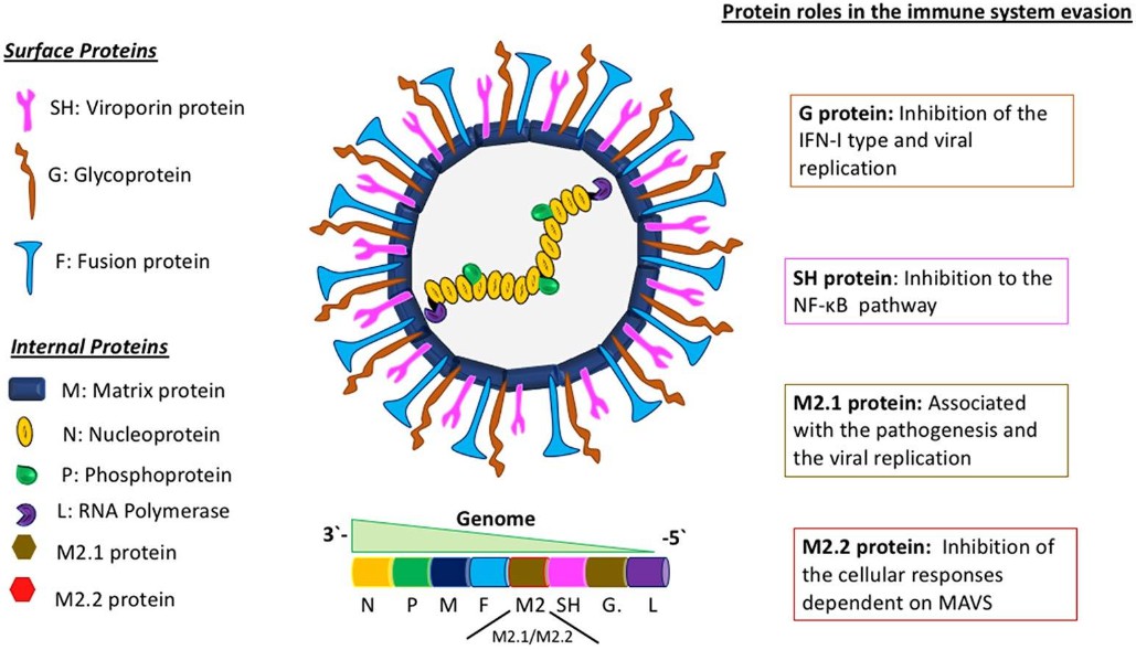 Figure 1. Schematic of the hMPV viral particle and its genomic organization.
(Source: Soto, J. A. et al., 2018)
HMPV enters the host cell by a viral host cell membrane fusion mechanism, in which F protein and G protein mainly mediate the adhesion of the virus to target cells and the fusion process between the virus and the cell membrane. Different from the RSV membrane fusion process, it has been reported that the replication level of recombinant hMPV lacking G protein and SH protein in cell culture medium is not affected, whereas G proteins might have an inhibitory effect on the fusion process mediated by F proteins of HMPV. This phenomenon suggests that F protein alone can mediate the membrane fusion process. This difference may be related to the lack of the Arg-Gly-Asp (RGD) sequence unique to HMPV F protein in RSV F protein.
The RGD sequence binds to integrins on the surface of host cells, especially αvβ1 integrin, to promote the adhesion process between viruses and host cells. During HMPV infection, the virus produces an inactive precursor protein (F0), which needs to be cleaved under the action of protease into two polypeptides F1 and F2 with membrane fusion activity connected by disulfide bonds. The fusion process mediated by the HMPV F protein is dependent on a low-pH environment and the histidine residues in the extracellular domain of the F protein have a great influence on its functionality. Experiments have shown that mutations in the histidine residue significantly reduce the protein's ability to promote cell fusion.
Previous studies have shown that the H435 residue in the HRB linking region plays the role of a pH sensor. When in a low pH environment, electrostatic repulsion between H435 and the surrounding region promotes the F protein-mediated membrane fusion process. Other studies have shown that MPRSS2 (a transmembrane serine protease) expressed in human lung epithelial cells can effectively promote the F protein cleavage process, providing favorable conditions for MPV infection and replication.
HMPV infects individuals through airborne droplets or direct contact. The virus first infects respiratory epithelial cells. The F protein and G protein on the surface of the virus bind to receptors on the host cell membrane and enter the cell. After that, the virus releases its genome, which serves as a template for the synthesis of viral proteins and the replication of viral RNA. The newly synthesized viral components assemble to form new viral particles, which are then released from the infected cell.
HMPV infection can lead to clinical symptoms affecting both the upper and lower respiratory tracts, with a higher prevalence in the latter. Common symptoms include coughing, bronchitis, bronchiolitis, and respiratory issues related to airflow obstruction. The immune response triggered by hMPV infection is characterized by a Th17-like response, involving the secretion of interleukin-6 (IL-6) and tumor necrosis factor-alpha (TNF-α) in the lungs.
In addition to the Th17-like response, an inadequate Th2-like immune profile is observed early in the infection, characterized by the secretion of IL-4, IL-5, IL-8, and other pro-inflammatory cytokines. Thymic stromal lymphopoietin (TSLP), a cytokine known to inhibit T cell activation, contributes to the delay in Th1-like immunity and promotes cytokine secretion associated with a Th2-like profile. This results in the excessive infiltration of neutrophils in the lungs and increased mucus production by goblet cells, leading to airway collapse.
Children under 2 years of age infected with hMPV are at a higher risk of developing asthma later in life, similar to the association observed with hRSV. Severe cases of hMPV infection can also contribute to chronic obstructive pulmonary disease (COPD) and exacerbate asthma symptoms in individuals. HMPV is considered one of the most prevalent viruses causing hospitalization in young children, accounting for about 10-12% of respiratory illnesses in children.
References
Loading ......
