
Inquiry Basket
 Loading ......
The dot blot is a simple and cost-effective technique used to detect and quantify specific proteins or nucleic acids in a sample. This technique has been widely used in research and diagnostics, and it is a popular alternative to more complex methods such as western blotting and ELISA. This technique involves the application of the sample directly to a membrane, which is then probed with a specific antibody or probe. The signal is then detected through a detection enzyme, and the intensity of the signal is proportional to the amount of target protein or nucleic acid in the sample.
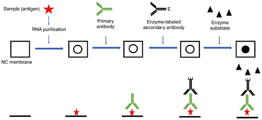 Fig. 1 Schematic diagram of dot blot technology.
(Zhu W,et al., 2019)
Before starting the dot blot assay, it is essential to gather all the necessary materials and reagents. These include:
The dot blot protocol involves several steps, including preparing the sample, preparing the membrane, setting up the dot blot apparatus, applying the sample to the membrane, blocking the membrane, incubating with the primary and secondary antibodies, and detecting the signal.
The first step in the dot blot protocol is to prepare the sample. This involves lysing the cells or tissue and extracting the protein or nucleic acid of interest. The sample should be diluted to a suitable concentration in a sample buffer. It is important to ensure that the sample buffer is compatible with downstream applications and that it does not interfere with the binding of the target protein or nucleic acid to the membrane.
Nitrocellulose or PVDF membranes are commonly used for dot blotting. These membranes have a high binding capacity and a low background signal. The membrane should be cut to the appropriate size to fit the dot blot apparatus. And draw a grid with a pencil to indicate the region you want to blot.
Use a narrow-mouth pipette tip to take 2 µL each of the standard dilutions at different concentrations, samples, negative control, and positive control and spot them on the square grid on the membrane. Minimize solution penetration area (usually 2-4 mm) by applying slowly. Care should be taken to avoid cross-contamination between samples. The samples should be allowed to dry completely before proceeding to the next step.
The fourth step in the dot blot protocol is to block the membrane to prevent the non-specific binding of primary and secondary antibodies. Blocking buffer is added to the membrane and incubated for a suitable amount of time (e.g., 1 hour at room temperature or overnight at 4°C). The blocking buffer should be compatible with downstream applications and should not interfere with primary and secondary antibody binding.
The primary antibody (0.1–10 µg/mL for purified antibody, 1:1,000–1:100,000 for antisera, 1:100–1:10,000 for hybridoma supernatant) specific to the target protein or nucleic acid is added to the membrane and incubated for a suitable amount of time in BSA/TBS-T. The membrane is then washed with washing buffer (TBS-T) three times (3 x 5 min) to remove any unbound primary antibody.
The secondary antibody conjugated to HRP is added to the membrane and incubated for a suitable amount of time. The membrane is then washed with TBS-T three times (1 x 15 min and 2 x 5 min), then once with TBS (5 min).
The substrate for the detection enzyme (e.g., ECL or BCIP/NBT) is added to the membrane, and the signal is visualized using an imaging system (e.g., chemiluminescence or colorimetric). The signal intensity is proportional to the amount of the target protein or nucleic acid in the sample.
The dot blot is a versatile technique with several applications. It can be used to quickly and easily confirm the presence of a target protein in a sample, as long as the primary antibody is specific to the target. Additionally, a dot blot can estimate the target protein concentration by comparing it to known control concentrations on the membrane. Dot blots can also be used to confirm antibody specificity, particularly for secondary antibody validation.
In addition, the dot blot protocol is commonly used to optimize primary and secondary antibody concentrations for Western blotting. During this process, the primary antibody is spotted directly onto the membrane and detected with the secondary antibody. To simplify the identification of the optimal primary: secondary concentration pair, the dot blot is often configured in a grid pattern.
The Creative Diagnostics team can provide you with the reagents you need for your spot-blotting experiments, including high-quality antibodies and assay kits to support your research. Whether you are a researcher or a diagnostician, a dot blot is a powerful tool to help you detect and quantify specific targets in your samples.
Reference
Loading ......
