
Inquiry Basket
 Loading ......
Streptococcus pneumoniae, commonly known as pneumococcus, is a pathogenic bacterium responsible for a range of infections, including pneumonia, meningitis, and otitis media. Understanding the structure and composition of its cell wall is crucial for understanding bacterial growth and cell-wall dynamics as well as developing effective diagnostic tools and therapeutic strategies.
The cell wall of Streptococcus pneumoniae is a complex structure composed of various macromolecules, including mesh-like peptidoglycan, teichoic acids, lipoteichoic acids, and surface proteins. These components work together to provide structural integrity and contribute to the pathogenicity of the bacteria.
Peptidoglycan
Peptidoglycan is a major component of the bacterial cell wall and forms a mesh-like structure surrounding the cell. It is composed of long chains of alternating N-acetylglucosamine (GlcNAc) and N-acetylmuramic acid ((MurNAc) residues, cross-linked by short peptide chains. The peptidoglycan layer inStreptococcus pneumoniae is unique and plays a crucial role in maintaining cell shape and protecting the bacterium from osmotic stress.
Wall Teichoic Acids
Wall teichoic acids (WTA) are another essential component of the pneumococcal cell wall. These long polymers are covalently attached to the peptidoglycan layer and extend outward from the cell surface. In Streptococcus pneumoniae, the structure of WTA is unusually complex, exhibiting the same repeating unit structure as lipoteichoic acid (LTA). Recent revisions have shed light on the intricacies of WTA structure, providing valuable insights into its role in pneumococcal pathogenesis.
Teichoic Acids
Teichoic acids in Streptococcus pneumoniae vary in length and can contain phosphorylcholine residues. Depending on the strain, teichoic acids may have an average length of 7-8 repeats and exhibit 1-2 phosphorylcholine residues per repeat. These modifications contribute to the bacterium's ability to evade the host immune system by mimicking host molecules or interfering with immune recognition. Understanding the structural variations in teichoic acids is crucial for developing targeted interventions against pneumococcal infections.
Surface Proteins
Streptococcus pneumoniae possesses a diverse array of surface proteins that are anchored to the cell wall. These proteins play crucial roles in various aspects of the bacterium's pathogenesis, including adhesion to host cells, evasion of the immune system, and biofilm formation. Surface proteins such as pneumococcal surface protein A (PspA) and choline-binding proteins (CBPs) have been extensively studied and are considered important virulence factors.
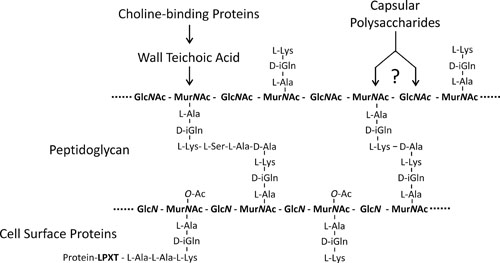 Fig. 1 Diagram of the cell wall complex of pneumococci. (Vollmer W, et al., 2019)
The regulation of bacterial cell-wall synthesis during growth and cell division is not well understood in pneumococci. Several proteins, including DivIVA, GpsB, StkP, and PhpP, play roles in this process. DivIVA is involved in cell pole formation and interacts with various proteins. GpsB contributes to peripheral and septal peptidoglycan synthesis, and its depletion leads to abnormal cell elongation and ring formation. The pneumococcal serine/threonine kinase StkP and its cognate phosphatase PhpP localize to division sites and are responsible for coordinating cell growth and division through reversible phosphorylation of cell division proteins. StkP may signal the status of the cell wall to other cell division proteins and regulate the switch from peripheral to septal peptidoglycan synthesis.
The cell wall of Streptococcus pneumoniae serves several essential functions that contribute to the bacterium's survival and pathogenicity.
Structural Integrity
The peptidoglycan layer, along with teichoic and lipoteichoic acids, provides structural integrity to the bacterial cell. It maintains cell shape and protects the bacterium from osmotic stress.
Host-Pathogen Interactions
The cell wall components of Streptococcus pneumoniae, particularly surface proteins, play a crucial role in the interaction between the bacterium and the host. Adhesion proteins facilitate the attachment of the bacterium to host cells, allowing it to colonize various tissues and cause infection. Other surface proteins help the bacterium evade the host immune system by interfering with immune cell function or modulating host immune responses.
Antibiotic Resistance
The cell wall of Streptococcus pneumoniae is a target for several antibiotics, such as beta-lactams. The bacteria have developed mechanisms to resist the action of these antibiotics by modifying the structure of the peptidoglycan layer or by producing enzymes that can degrade antibiotics. Understanding the mechanisms of antibiotic resistance in the cell wall is crucial for the development of new therapeutic strategies.
In conclusion, understanding the composition and functions of the cell wall is essential for developing diagnostic tools and therapeutic strategies to combat pneumococcal infections. As a leading company in the field of biotechnology, Creative Diagnostics is dedicated to advancing research in this area and developing innovative solutions for the detection and treatment of Streptococcus pneumoniae infections.
References
Loading ......
