Materials of Single-Cell Western Blotting Protocol
- Regular microscope slide.
- Photoactive polyacrylamide gel.
- SU-8 2025 micropost.
- 2× 8-well microarray hybridization cassette.
- Radio immunoprecipitation assay (RIPA) lysis/electrophoresis buffer—0.5 % SDS, 96 mM glycine, 0.1 % v/v Triton X-100, 0.25 % sodium deoxycholate 0.25 % sodium deoxycholate in 12.5 mM Tris, bring to pH 8.3. RIPA buffer is denaturing but nonreducing.
- UV mercury arc lamp (Lightningcure LC5 Hamamatsu).
- Gel precursor solution—8 % T (w/v total acrylamide), 2.7 % C (w/w of the cross-linker N, N -methylene bisacrylamide) from 30 % T, 2.7 % C stock, 3 mM BPMAC from 100 mM stock in DMSO, 0.1 % SDS, 0.1 % Triton X-100, 0.0006 % riboflavin 5′ phosphate, 0.015 % ammonium persulfate (APS), 0.05 % tetramethylethylenediamine (TEMED) in 75 mM Tris buffer. Titrate with HCl to a pH of 8.8.
- Dichlorodimethylsilane (DCDMS).
- Phosphate-buffered saline (PBS)—pH 7.4.
- SU-8 developer solution (Microchem Corp).
- TBST—Titrate 100 mM Tris, containing 150 mM NaCl, with HCl to pH 7.5, followed by 0.1 % Tween 20.
- Bovine serum albumin (BSA).
- Stripping buffer—2.5 % SDS, 1 % β-mercaptoethanol, 62.5 mM Tris. Titrate to a pH of 6.8 with HCl.
- Platinum wire electrodes (0.5 nm diameter, Sigma-Aldrich).
Methods of Single-Cell Western Blotting Protocol
Method of Culturing Cells (See Note 3)
- Neural stem cells are extracted from hippocampus of an adult rat.
- Coat the culture-treated polystyrene plates with polyornithine 10 μg/mL and laminin 5 μg/mL.
- Culture the cell in required media. Preparation of media: Take 250 mL of DMEM-F12 and mix with 250 mL N-2. Add 10 μg of recombinant human FGF-2 (Peprotech). Culture up to 80 % of confluency.
- For cell detachment use accutase.
Preparation of Protein Standard (Ladder)
For making protein standard or ladder for single-cell western some of the fluorescent antibodies are prepared, and others are obtained in premade condition. The ladder is made by purified proteins having molecular weight of 27–132 kDa. The following proteins can be used:
- Purified Dronpa—27 kDa
- OVA—ovalbumin—45 kDa
- BSA—66 kDa
- OVA dimer—90 kDa
- BSA dimer—132 kDa
Purified albumin and BSA were Alexa Fluor 488 labeled and can be obtained from Life Technologies. Purified dronpa can be prepared by expression in Rosetta-competent cells transformed with tobacco etch virus (TEV) ligase-independent cloning vector and then affinity purified.
 Figure 1. Construction of protein ladder.
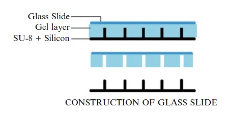 Figure 2. Construction of glass slide.
The fluorescence at different distance is recorded and the distance measured. The fluorescence within 10 % of the target region is considered as positive.
Preparation of Fabricated Slides
- Microposts (SU-8) are fabricated on silicon wafer by standard lithography method.
- Spin SU-8 2025 according to the manufacturer's guidelines to reach a thickness of 30 μm.
-
Expose it to UV light (365 nm) at around 40 mW/cm2.
- Arrange the features in square configuration. Keep the pitch as follows:
(a) 500 μm in direction of separation.
(b) 190 μm in transverse direction.
(c) 2 × 8 blocks of 14 × 30 are separated by space of 9 mm in order to accurately match the dimensions of 2 × 8-well microarray hybridization cassette. - Optical profilometry can be done to analyze the uniformity of thickness using SU-8 developer solution.
- Silanize the wafer by using vapor deposition of DCDMS for approximately 1 h in vacuum.
- Wash the wafer with deionized water and dry it using nitrogen stream.
- In the next step, take a microscope glass slide and silanize it in order to create a surface layer of methacrylate functional group.
- Silanized slides are placed on micropost wafer and aligned to SU-8 rail and micropost features.
- Prepare gel precursor solution (as described in materials).
- Sonicate the precursor solution and degas it for 1 min in vacuum before addition of Triton X-100, SDS, APS, riboflavin, and TEMED.
- Add the precursor solution in the area between glass slide and silicon wafer with the help of pipette.
- Wait for 30 s for the solution to settle.
- Expose the glass slide to blue light for 7.5 min at 4,701× from a collimated 470 nm LED.
- Leave it for 10–11 min for polymerization of gel.
- Wet the edges of the fabricated slide with 1–2 mL PBS.
- Carefully separate the slide from wafer with the help of blade.
- Fabricated slides are now ready for experiment. These slides can be stored at 4 °C in PBS for 1–2 weeks.
Single-Cell Western Blot
- Remove the slides from PBS solution.
- Tilt the slide to one side and drain the excess liquid from the corner of the slide. Absorb it with the help of Kimwipe.
- Apply 1–2 mL of cell suspension solution and wait for settling of cells.
- Place the slide in petri dish 100 × 100 mm.
- After every 2–5 min gently shake the petri dish for 10 s.
- The settling time ranges from 5 to 30 min.
- Observe the settling of cells and microwell occupancy under bright-fi eld microscope.
- After cells are settled down in microwells, lift one end of the slide approximately at an angle of 10–20° for removal of excess media. Pipette 1 mL of PBS to the raised side of the slide 4–5 times in order to remove the cells on the surface of the slide. Use vacuum at low pressure if necessary.
- For cell counting, put 1 mL of PBS on the slide and place another covering slide to prevent bubble formation.
- Image under bright-fi eld microscopy at 4× magnification controlled by MetaMorph software.
- After the counting is done, remove the upper glass slide gently across the gel layer.
- Transfer the slide with settled cell to 60 × 100 mm customized horizontal electrophoresis chamber fabricated from 3 mm plastic (Perspex).
- Place platinum wire electrode along the long edge of the chamber and connect it to standard electrophoresis power supply with the help of alligator clips.
- Attach the slides to the bottom of the chamber using petroleum jelly.
- Add 10 mL of RIPA lysis buffer to the slide to lyse the cells and wait for 10 s.
- Turn on the current with setting of 200 V (E = 40 V/cm) for about 30 s.
- Monitor the separation of proteins from a single cell in real time with a magnification of 10×. The filters are optimized for particular cells and may vary in different cell types.
-
After separation is done, expose to UV light using UV mercury arc lamp at a distance of 10 cm above the slide with a UW power of 40 mW/cm2. The exposure helps in immobilization of proteins.
- Wash the slides with 10 mL of denaturing RIPA buffer for about 10 min.
- Wash the slides with 10 mL of TBST for about 10 min (at this point the slides can be stored up to a week at 4 °C in TBST).
- Incubate each block of separation with 40 μL of primary antibody solution diluted in TBST and 2 % BSA.
- Keep the slide on rotator for about an hour under gentle shaking mode.
- Remove the slides and wash the slide adding 10 mL of TBST and place on the rotator for 15 min under gentle shaking mode.
- Repeat the above step two times for a total of three washes or 45 min.
- Incubate each block of separation with 40 μL of fluorescently labeled secondary antibody solution diluted in TBST and 2 % BSA.
- Remove the slides and wash the slide adding 10 mL of TBST and place on the rotator for 15 min under gentle shaking mode. Repeat the same steps two times for a total of three washes or 45 min.
- Finally wash the slide with 10 mL of deionized water for 5 min and dry under nitrogen.
- The slide is ready for imaging studies. Use microarray scanner for imaging studies.
Single-Cell Western Blot for Purified Protein
- The same protocol is used for purified proteins with few different steps.
- Slides are incubated in RIPA denaturing buffer for 30 min and then submerged in fresh RIPA buffer for about 5 s.
- A second glass slide is placed to trap the proteins.
- The sandwiched slide is then subjected to electrophoresis and photo-capture using UV light. Second glass cover can then be removed.
- The remaining steps are similar.
Reprobing with Another Antibody
- Unlike conventional western blotting, the slide of single-cell western blot can be reused again for detection of another protein.
- Heat the stripping buffer to 50 °C.
- Apply the stripping buffer to slide and incubate for 3 h.
- After the stripping is done, wash the slides with 10 mL of TBST for about 5 min three times.
- The slides are ready for the next set of primary and secondary antibodies.
- Air-dried slides can be reused again when preserved at 4 °C.
Analysis of Single-Cell Western Blot
Analysis of single-cell western blot involves confirmation of presence of single cell in a well. Well containing no cell or more than one cells is not included. The distance between the fluorescent purified protein and well is determined. The fluorescence activity within 10 % of the region can be considered on-target.

 Loading ......
