
Inquiry Basket
 Loading ......
Apoptosis, or programmed cell death, is a crucial physiological process that plays a fundamental role in various biological phenomena, including embryonic development, tissue homeostasis, and immune response. At the molecular level, apoptosis is tightly regulated by a family of enzymes known as caspases. Caspases are cysteine proteases that orchestrate the sequential events leading to cell death.
Caspases are categorized into two main groups based on their function in apoptosis: initiator caspases and executioner caspases. Initiator caspases, including caspase-2, -8, -9, and -10, are primarily responsible for initiating the apoptotic process. They are activated in response to various apoptotic stimuli, such as DNA damage, death receptor signaling, or mitochondrial dysfunction. Once activated, initiator caspases cleave and activate downstream executioner caspases, amplifying the apoptotic signal.
Executioner caspases, including caspase-3, caspase-6, and caspase-7. They are activated by initiator caspases and are responsible for the proteolytic cleavage of numerous cellular substrates, leading to DNA fragmentation, cytoskeletal reorganization, and ultimately, cell death. Caspase-3, in particular, is considered a key executioner caspase and is widely used as a marker for apoptotic cell death.
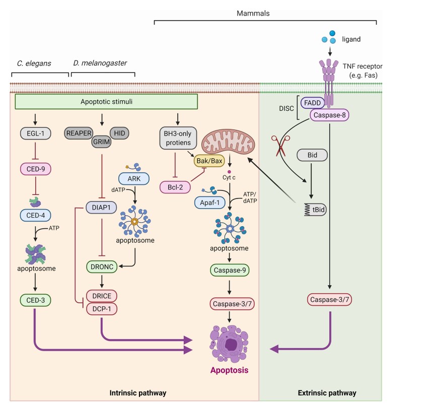 Fig. 1 Caspases are evolutionarily conserved effectors of apoptosis. (Kumar S, et al, 2022)
Caspase activation is a sequential multi-step hydrolysis process. First, the N-terminal propeptide is hydrolyzed at a specific site between the N-terminal propeptide and the large subunit of the caspase precursor, and then the large and small subunits are released by cleaving between the large and small subunits. The subunits form heterodimers, and the two dimmers form an active tetramer.
Removal of the N-terminal propeptide is the first step and necessary for the activation of caspase, but activation of caspase-9 does not require removal of the N-terminal propeptide. There are basically two mechanisms for caspase activation, namely homologous activation and heterologous activation, which are closely related. Caspases that undergo homologous activation are also called initiator caspases. After inducing apoptosis, the initiating caspases are recruited to specific initiation activation complexes through adapters, forming homologous dimer conformational changes that lead to enzymatic cleavage between homologous molecules and self-activation. Typically, caspase-8, -10, and -2 mediate cell apoptosis through the death receptor pathway, and are recruited to Fas and TNFR1 death receptor complexes, respectively. Caspase-9 participates in cell apoptosis through the mitochondrial pathway and is recruited into apoptosomes composed of Cyt c/d ATP/Apaf-1.
The initiation of caspase activation initiates the intracellular death program, which amplifies the apoptotic signal by hydrolyzing downstream caspases through hetero-activation and simultaneously transmits the death signal downstream. Heterologously activated caspases are also known as executioner caspases. Unlike initiator caspases, executioner caspases cannot be recruited or bound to the initiation complex, and they are dependent on initiator caspases for activation.
Caspase activation is tightly regulated, and two important regulatory mechanisms are mediated by FLIP and inhibitors of apoptosis proteins (IAPs).
FLIP, both viral (v-FLIP) and cellular (cFLIP), inhibits caspase-8 activation by interacting with FADD, thereby interfering with apoptosis signaling through death receptors. Upregulation of cFLIP by the NF-κB pathway promotes cell survival in response to TNF. However, TNFα-mediated JNK activation can lead to the degradation of FLIP, sensitizing cells to apoptosis.
Mammalian IAP-like proteins, including XIAP, cIAP-1, cIAP-2, NAIP, ML-IAP, ILP-2, and surviving. Some of them can interact with caspases and inhibit mature caspase-3, -7, and -9. Among them, XIAP is the primary inhibitor at physiological concentrations. Structural studies reveal that the N-terminal BIR2 linker region of XIAP occupies the catalytic site of active caspase-3 and -7, while the BIR2 and BIR3 domains are involved in inhibiting caspase-9. During apoptotic signaling, SMAC/DIABLO and HtrA2 can disrupt XIAP's inhibitory effect by binding to its BIR domains, thus allowing caspase activity.
Antibody-based methods and substrate-based methods are commonly used to detect and measure caspase activity.
Antibody-based methods rely on the use of specific antibodies that recognize and bind to active caspases. The choice of antibody is crucial as it determines the specificity and location of the targeted epitope. By selecting the appropriate antibody, researchers can accurately detect active caspases in their experimental systems.
Substrate-based methods utilize biochemical substrates consisting of short peptides with specific cleavage sequences recognized by caspases. These peptides are covalently attached to colorimetric or fluorogenic detection probes. When caspase leaves the substrate, the attached compound is released, resulting in an increase in absorbance (colorimetric substrate) or fluorescence (fluorogenic substrate). The magnitude of the signal corresponds to the amount of caspase activity present in the sample.
In conclusion, understanding the role of caspases in apoptosis provides valuable insights into the mechanisms underlying various physiological and pathological processes. As a leading company in the field, Creative Diagnostics is dedicated to advancing the understanding of apoptosis and developing innovative tools for caspase research, facilitating progress in biomedical research and therapeutic interventions.
References
Loading ......
