
Inquiry Basket
 Loading ......
Gallbladder cancer ranks first among malignant tumors of the gallbladder, followed by sarcomas, carcinoids, primary malignant melanoma, giant cell adenocarcinoma, and others. Primary gallbladder cancer is relatively rare in clinical practice, accounting for only about 1% of all cancers according to domestic reports. Gallbladder cancer is a relatively rare but aggressive form of gastrointestinal malignancy that arises from the epithelial cells lining the gallbladder. Due to its nonspecific early symptoms and anatomical location, gallbladder cancer is often diagnosed at an advanced stage, contributing to its poor prognosis.
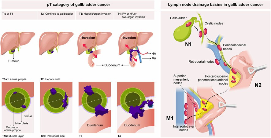 Figure 1. Staging system for gallbladder cancer. (Zhou, Yanzhao, et al. 2023)
Gallbladder cancer exhibits significant geographic and demographic variations worldwide. It is considered the sixth most common gastrointestinal cancer, with approximately 220,000 new cases diagnosed globally each year, according to recent data from the World Health Organization (WHO).
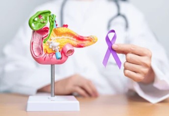
Regional Distribution: The highest incidence rates are observed in certain regions of South America (such as Chile), Central Europe, and Asia (particularly India and Pakistan). For example, in parts of northern India, the incidence can be as high as 20 per 100,000 population, which is significantly higher than the global average of 3 per 100,000. In contrast, North America and most of Western Europe report lower rates, typically below 2 per 100,000.
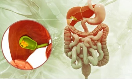
Demographic Factors: Gallbladder cancer affects women more frequently than men, with a female-to-male ratio of approximately 3:1. This gender disparity is thought to be linked to the higher prevalence of gallstones in women, a key risk factor. The disease is most commonly diagnosed in individuals aged 60 to 70 years, although cases can occur in younger adults, especially those with underlying risk factors.
The pathogenesis of cancer is complex, and numerous genetic and epigenetic events are necessary for this process. Malignancy and invasiveness of cancer are significantly associated with mutations and functional defect in hMLH1 and hMSH2 genes that function as part of mismatch repair (MMR) mechanism. FHIT gene in chromosome 3p14.2 is downregulated and partially deleted with increased tumor grading and lymph node metastasis. Telomerase is an enzyme which has the property of immortality and high expression of it is seen in gallbladder cancer tissue. Matrix metalloproteinases (MMPs) such as MMP-2 are also upregulated and involved in tumor invasion and metastasis, and the level of MMP inhibitors such as TIMP-2 can act as a prognostic marker for the disease. COX-2, E-Cadherin, i-NOS, and VEGF overexpression is associated with worse prognosis and advanced stage of the disease.
1. Ultrasound examination
Ultrasound examination is simple, non-invasive, and can be used repeatedly, making it the preferred method of examination. Endoscopic ultrasound using high-frequency probes to scan the gallbladder only through the stomach or duodenal wall significantly improves the detection rate of gallbladder cancer and can further determine the degree of tumor infiltration in each layer of the gallbladder wall structure.
2. CT scan
The imaging changes of gallbladder cancer in CT scan can be divided into three types:
(1) Thickened gallbladder wall with localized or diffuse irregular thickening;
(2) Nodular papillary nodules protrude from the gallbladder wall into the gallbladder cavity;
(3) The solid form is formed by the extensive infiltration and thickening of the gallbladder wall by tumors, combined with the filling of intracavitary cancer blocks to form a substantial mass. If the tumor invades the liver or metastasizes to the lymph nodes of the hepatic hilum and pancreatic head, it can often be displayed under CT imaging
3. Color Doppler flow imaging
The gallbladder mass and abnormal high-speed arterial blood flow signals detected within the wall are important features that distinguish primary malignant tumors of the gallbladder from metastatic or benign gallbladder masses
4.ERCP
The diagnostic rate of ERCP for gallbladder cancer that can display the gallbladder can reach 73% to 90%. But more than half of ERCP examinations cannot show the gallbladder.
5. Cytological examination
(1) Cytological examination can directly take biopsy or extract bile to search for cancer cells. The positive rate of cytological examination is not high, but combined with imaging examination, more than half of gallbladder cancer patients can still be diagnosed.
(2) The positive rate of CEA in gallbladder cancer was 100% in the CEA immunohistochemical study report of tumor markers in tumor specimens. The serum CEA level in patients with advanced gallbladder cancer can reach 9.6ng/ml, but it has no value in early diagnosis. CA19-9, CA125, CA15-3 and other tumor glycan antigens can only be used as auxiliary examinations for gallbladder cancer.
Radical surgery is the first choice of definitive treatment for gallbladder cancer, and it is the only means of curing gallbladder cancer. In the treatment of advanced gallbladder cancer patients who are not suitable for curative resection and cannot tolerate surgery, the treatment adopts palliative therapy, for the purpose of relieving the symptoms of gallbladder and biliary tract infection, improve liver function, and increase the quality of life.
| Treatment | Descrpition |
| Surgery | The surgical method is selected according to the stage of tumor discovery. For early gallbladder cancer without lymph node and other site metastasis, cholecystectomy can be performed, that is, the gallbladder is surgically removed. For middle gallbladder cancer, the scope of surgery resection should be extended. In addition to the removal of the gallbladder, partial liver and hepatoduodenal ligament lymph nodes should also be removed. For advanced gallbladder cancer, if the physical condition permits, extended radical surgery for gallbladder cancer can be performed. The range of resection is larger, the trauma is more, and the risk of complications is higher |
| Chemotherapy | There is no recommended and unified chemotherapy regimen for gallbladder cancer at present. It has been confirmed by research that adjuvant chemotherapy after radical surgery for gallbladder cancer can improve survival. |
| Radiotherapy | The radiotherapy of gallbladder cancer includes preoperative radiotherapy, intraoperative radiotherapy, intracavitary radiotherapy and non-surgical palliative radiotherapy. Gallbladder cancer patients with early stage but with lymph node metastasis are suitable for radiotherapy. |
| Interventional therapy | Patients with gallbladder cancer with extensive metastasis and lost surgical opportunity can be treated with biliary drainage to relieve symptoms and improve quality of life. |
Gallbladder cancer remains a highly lethal malignancy due to late diagnosis and aggressive biology. While surgical resection offers the best chance for cure in early stages, advanced disease requires a multidisciplinary approach involving chemotherapy, radiation, and emerging therapies. Increased awareness, early intervention for gallstone disease, and research into novel treatments are crucial for improving outcomes.
Reference
| Cat. No. | Product Name | Host | Isotype | Application | |
| DCABH-200291 | Anti-KIAA0825 monoclonal antibody | Rabbit | IgG | WB, ELISA | Inquiry |
| CABT-B1139 | Anti-ABCB11 (Cytoplasmic domain) monoclonal antibody, clone 20E5.2 | Mouse | IgG1, κ | WB, IHC-P | Inquiry |
| DPABH-22846 | Anti-ABCG8 (aa 299-415) polyclonal antibody | Rabbit | IgG | ICC/IF, WB, IHC-P | Inquiry |
| Cat. No. | Product Name | Size | Species Reactivity | Application | Detection Method | |
| DEIA1977 | Fasciola IgG ELISA Kit | 96T | Human | Qualitative | iELISA | Inquiry |
| Cat. No. | Product Name | Size | Target | Species | |
| DAGA-805 | CA 50 | 50 kU | / | / | Inquiry |
| CDBP0719 | Mouse CCKAR blocking peptide | 100 g | CCKA Receptor (mouse) | Mouse | Inquiry |
| CDBP0720 | Human CCKAR blocking peptide | 100 g | CCKAR | Human | Inquiry |
| DAG-WT1206 | Recombinant Canine Pancreatic Lipase | 1 mg | PNLIP | / | Inquiry |
Loading ......
