
Inquiry Basket
 Loading ......
Enzymatic immunohistochemistry, as the name implies, utilizes enzyme-labeled antibody-antigen reactions is histologic tissue sections to enable the visualization of specific cellular constituents or antigens present on cellular elements. The final step in the reaction utilizes a chromogen, which when cleaved by the enzyme-labeled antibody produces a stable, colored compound that can be observed with the aid of a light microscope. Direct detection techniques utilize a one-step staining method that involve an enzyme-labeled antibody reacting directly with the antigen in tissue sections. Although quick to perform, it lacks sensitivity and it is therefore rarely used in current practice. The indirect method involves an initial reaction with an unlabeled primary antibody. In the next step, a second labeled antibody reacts with the primary unlabeled antibody leading to an amplified signal. The most commonly employed enzyme labels include horseradish peroxidase (HRP), alkaline phosphatase (AP) or glucose oxidase. The addition of a chromogen serves as the last steps in the procedure. The enzyme present on the antibody reacts with the chromogen to yield an insoluble, colored compound. As further developments occurred in this field, newer detection systems came to exist. The PAP (peroxidase–anti-peroxidase), the avidin–biotin complex (ABC), and labeled streptavidin–biotin (LSAB) methods proved more sensitive than the older one- or two-layer techniques. These next generation IHC methods emerged in 1981 and are still being used today.
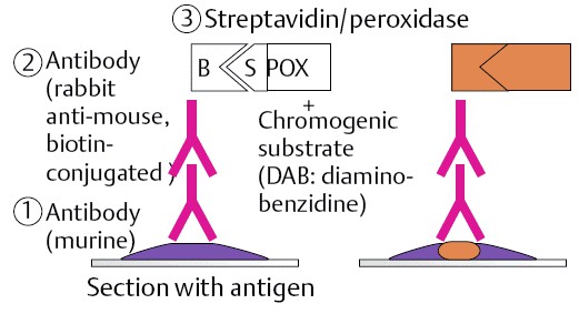 Figure 1. The principle of Immunohistochemistry (IHC) enzyme labeled technique. (Basma Fathi Alanbari, et al.;2016)
A third layer is involved in the PAP method, which is a rabbit antibody to peroxidase, coupled with peroxidase to produce a very stable peroxidase–anti-peroxidase complex. The avidin–biotin complex (ABC) method relies on the strong affinity of Avidin (derived from chicken egg) for the vitamin biotin, which is easily conjugated to antibodies and enzymes. In this method, secondary antibodies are conjugated to biotin and function as links between tissue-bound primary antibodies and the avidin–biotin–peroxidase complex. The labeled streptavidin–biotin (LSAB) method also utilizes a biotinylated secondary antibody that joins primary antibodies to a streptavidin–peroxidase conjugate. The advantage of this method over the ABC method is that in contrast to avidin (a glycoprotein with an isoelectric point of 10, which binds nonspecifically to lectin-like and negatively charged tissue components at physiologic pH), streptavidin has a more neutral isoelectric point and lacks the carbohydrate moieties, resulting in less non-specific binding.
In this chapter, we will utilize histologic sections derived from formalin-fixed, paraffin-embedded (FFPE) tissue to identify antigens using horseradish peroxidase or alkaline phosphatase-labeled antibodies.
Unless a different temperature is specified, perform all steps at room temperature. All incubations should be carried out in a humidified chamber to avoid drying of the tissue. Always wear personal protective equipment (laboratory coat, safety glasses, and gloves) when handling reagents and glass slides.
Implementation of this procedure implies that appropriate slide preparation has been carried out ahead of time, including the removal of paraffin from tissue sections, tissue rehydration as well as unmasking of the antigen (antigen retrieval). Optimal dilution of and incubation times should be determined for each primary antibody prior to use. A positive control tissue is strongly recommended to ensure that the antibody is performing as expected. It may also be useful to include a negative control; to do so, simply replace the primary antibody with antibody diluent.
References
Loading ......
