Introduction of ELISPOT to Study Microglia's TNFα
The ELISPOT assay was originally designed to study the secretion of antibodies from B cells and then was modified to study secretion of cytokines and chemokines from immune system cells. Due to its high sensitivity, ELISPOT remains the technique of choice to study the frequency of cytokine secreting immune system cells in cancer research, AIDS research, and immunization and vaccine studies.
ELISPOT assays are typically used to study cytokine secretion by cells from peripheral blood and lymphoid tissues. However, this technology can be used on other cell types, including microglial cells in the central and peripheral nervous systems. Microglial cells are the resident macrophages in brain and in spinal cord and provide immune defense of the CNS: they resemble immune system cells in that they also secrete cytokines and chemokines. For example, microglia may secrete pro-inflammatory cytokines, including IL-1β and TNFα, that can affect neuronal function, induce neurodegeneration, and are also implicated in pain.
In our current study, we adapted the ELISPOT assay to study the frequency of TNFα secreting microglial BV2 cells, which are similar to naturally occurring microglial cells. Our study has shown that, in addition to immunology studies, ELISPOT can also be used as a powerful tool for neuroscience research to study mechanisms underlying protein secretion from microglial cells. In addition, ELISPOT can be used for high-throughput screening of potent neuroprotective drugs.
Methods of ELISPOT to Study Microglia's TNFα
Culture of BV2 Cells
Like many other types of cells, BV2 cells are stored frozen in liquid nitrogen (e.g., five million cells per vial) and before they can be plated into the ELISPOT plates they need to be thawed and transferred into fresh culture media. Procedures are performed at room temperature unless specified otherwise.
- Remove a vial with frozen BV2 cells from liquid nitrogen storage tank and thaw it rapidly at 37°C (1 min/1 mL of cell suspension).
- Add 1 mL of thawed cells to 15 mL growth media prewarmed at 37°C.
- Spin cells in a centrifuge at 500 × g for 5 min at room temperature.
- Remove supernatant and discard it. Add 10 mL of growth media.
- Spin cells again in a centrifuge for 5 min.
- Discard supernatant and resuspend cells in 15 mL of a fresh growth media in a culture flask.
- Culture cells until they are confluent (approximately 48 h).
- Prewarm fresh culture media at 37°C.
- Discard the media from the culture flask and wash adhered BV2 cells twice with 10 mL of PBS.
- Add PBS/EDTA to the flask and incubate at 37°C until cells begin to detach (10–15 min).
- Shake cells, add 15 mL of growth media, and transfer cells into a 50-mL conical tube.
- Centrifuge cells for 5 min at 500 × g.
- Discard supernatant and resuspend cells in 10 mL of prewarmed growth media.
- Mix cell sample (50 ML) 1:2 with trypan blue dye and pipette 10 ML of that mixture into each side of a hemacytometer under a coverslip. Count cells under the microscope using a 20× lens and phase contrast condenser (see Note 1).
ELISPOT Assay
- Plate BV2 cells (100 ML/well) into the ELISPOT plates at cell concentrations of 1.2 ×10³ and 104 cells/mL (see Note 2).
- Stimulate BV2 cells with LPS at 1 Mg added directly to cells in ELISPOT plates and incubated in a CO2 incubator at 37°C for 18 h (see Notes 3–5).
- After finishing the incubation, remove growth media from the plates with a multichannel pipette and wash the plate once with 1× PBS and then add 100μL of PBS/EDTA to plate for 10 min to remove adherent cells. Wash the plate by rinsing wells four times with wash buffer (see Notes 6–8).
- Make working solutions of the detection antibodies by mixing concentrated detection antibodies 1:120 with dilution buffer.
- Add 100μL of detection antibody working solution into each well and incubate ELISPOT plates overnight in the refrigerator or in the cold room at 2–8°C (see Note 9).
- Wash plates three times with the wash buffer.
- Prepare a working solution of streptavidin-alkaline phosphatase by mixing the concentrated stock solution 1:120 with the corresponding dilution buffer.
- Add 100 μL of streptavidin-alkaline phosphatase working solution to each well and incubate for 2 h.
- Wash plates three times with wash buffer.
- Add 100 μL of ready-to-use BCIP/NBT substrate into each well and incubate for 30–60 min in a place protected from direct light.
- Wash plates three times with distilled water and let them dry completely.
- Quantify spots using an automated ELISPOT reader (see Note 10).
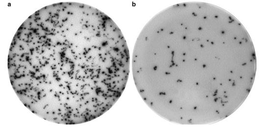 Figure 1. Typical ELISPOT image of TNFA secreted by microglial BV2 cells.