Medica 2026
Nov 16-19, 2026 - Düsseldorf, Germany
ADLM 2026
July 26-30, 2026 – Anaheim, CA, USA

Double-Sandwich ELISA Protocol

Introduction of Double-Sandwich ELISA Protocol

The enzyme-linked immunosorbent assay (ELISA) has enjoyed application in many areas because of its high specificity and sensitivity. Many variations are known including the indirect ELISA, competitive ELISA, the antibody-sandwich ELISA, antibody-capture ELISA, and the double antibody-sandwich ELISA. One of the most useful immunoassay formats is the sandwich ELISA designed for detection of soluble antigens. The difference between a capture ELISA and a sandwich ELISA is that a capture ELISA is designed to measure the amount of antibody present in a sample (typically a serological test), while a sandwich ELISA measures the amount of antigen in the sample. Thus, for a sandwich ELISA, a pair of antibodies to the target antigen is needed. The antigen is trapped between the capture antibody and the detector antibody. The example shown represents a direct binding sandwich ELISA since the detector antibody is directly labeled with an enzyme. A common variant is the indirect sandwich ELISA where the binding of the detector antibody is visualized by binding of a third antibody that is conjugated to the reporter molecule (i.e., if the detector antibody is made in a goat, the third or “reporter antibody” would be an enzyme-conjugated anti-goat antibody). In this scenario, the primary capture antibody could not be produced in goats but could be a mouse monoclonal antibody.

The detector and/or reporter antibody can be labeled with an enzyme and the sandwich detected with suitable colorimetric, fluorescent, or luminescent substrates. Alternatively, the detector antibody can be labeled with biotin and the sandwich detected using enzyme-conjugated streptavidin. The basic requirement of a sandwich ELISA (unless a repetitive epitope exists on the target antigen) is that two antibodies binding different epitopes on the same antigen are required. Furthermore, binding of one antibody must not interfere with binding of the second antibody even if they bind different epitopes.

Schematic of typical sandwich ELISAs. 
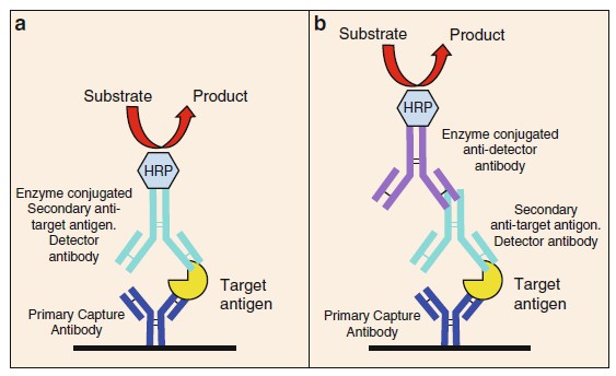
Figure 1. Schematic of typical sandwich ELISAs.

Since the performance characteristics (specificity and sensitivity) of a sandwich ELISA are directly related to the quality of the antibodies (their binding affinities, stability, etc.). Finding matched antibody pairs, especially two monoclonal antibodies (mAbs), with these properties is often difficult. Monoclonal antibodies offer many advantages versus polyclonal antibodies including identification of the antibody-binding epitope, improved assay specificity and reliability since a positive result requires binding of two highly specific reagents with known epitopes, the ability to select high- affinity conformational antibodies with the desired binding specificity, and a consistent source of high-quality reagent. The sandwich ELISA itself has many advantages including that the sample need not be purified before analysis. Antigen purification and concentration is accomplished during the capture phase of the assay resulting in a three- to five fold (or greater) increase in sensitivity versus a direct or indirect ELISA. Sandwich ELISAs usually display high specificity and greater confidence in the result because target detection requires binding of two distinct antibodies. However, their ability to detect low levels of target in complex samples makes them ideal tests for measuring the presence of the target antigen in unknown samples, e.g., food, environmental, or clinical samples.

Research in our laboratory has focused on development of sensitive immunoassays for botulinum neurotoxin (BoNT). Botulinum neurotoxins are produced by the anaerobic bacterium Clostridium botulinum. These toxins, as with many bacterial toxins, are large, complex di-chain protein toxins. In the case of BoNT, the active molecule consists of a 100 kDa heavy chain and a 50 kDa light chain joined by a single disulfide bond. Our initial approach was to screen hybridoma cell fusion supernatants using traditional indirect ELISA. Neurotoxin was adsorbed onto the bottoms of 96-well microassay plates. Supernatants from cell fusion experiments were added and antibody binding detected using an anti-mouse antibody conjugated to horseradish peroxidase (HRP). Earlier studies to develop monoclonal antibody-based sandwich ELISA for detecting BoNT serotype B were only partially successful since the majority of the mAbs isolated failed to bind the toxin in solution. All of the mAbs isolated displayed strong binding in the indirect ELISA (impressive titration curves), good specificity (i.e., binding serotype B but not serotype A), and strong binding on Western blots. However, none of these antibodies bound the toxin in solution; hence, none were suitable as capture antibodies for a sandwich ELISA. A discouraging result after expending significant resources to generate the hybridoma clones, purify the mAbs and label with biotin in order to identify suitable antibody pairs for a sandwich ELISA. Adsorbing protein antigens onto the plastic surface of microassay wells can result in modifications of the protein. Many changes can be envisioned including changes to the surface charge of the protein, mild denaturation resulting in exposure of cryptic epitopes, and blocking surface epitopes. Studies in our laboratory suggested that BoNT was modified when absorbed onto the plastic bottoms of microassay wells. Similar results were observed with two different BoNT serotypes and with nontoxic BoNT-associated proteins. Interestingly, the BoNT antibodies could be induced to bind toxin in solution by treating the toxin with weak SDS solutions or adjusting the pH. However, these steps necessitate additional sample preparation and only marginally improved assay performance. The double-capture ELISA outlined was applied as a screening tool to evaluate large numbers of hybridomas supernatants following cell fusion experiments in order to select for mAbs able to capture toxin in solution. Using this screen, it has been possible to select useful antibody pairs and construct sandwich ELISAs for both BoNT serotypes B, E, and F (latter two unpublished) as well as good antibody pairs for the nontoxic BoNT hemagglutinin-70-associated protein.

Once mAbs suitable for a sandwich immunoassay are identified, they can be formatted into numerous different tests, e.g., sandwich ELISA, lateral-flow devices, and various immunobiosensors. The critical reagents in these different assay formats are the antibodies.

Materials of Double-Sandwich ELISA Protocol

Prepare all solutions using ultrapure water (prepared by reverse osmosis to attain a sensitivity of 18 MΩ at 22 °C). All reagents were stored at 4 °C (unless indicated) and warmed to room temperature before use. All studies involving animals followed protocols approved by the USDA Western Regional Research Center’s Institutional Animal Care and Use Committee.

Double-Capture ELISA Components

  1. Tris-buffered saline (TBS): 0.2 M Tris–HCl, 0.9 % NaCl, pH 7.4. Add 900 mL water to a 1-L bottle. Add 100 mL of a 10× Tris-buffered saline solution. Mix and store at 4 °C.
  2. TBS containing 0.05 % Tween-20 (TBST). Adjust 0.5 mL of Tween-20 to 1 L with TBS.
  3. 0.05 M carbonate buffer (pH 9.6). Dissolve one buffer capsule in 100 mL water.
  4. Blocking solution: 5 % powered nonfat milk in TBST. Store at 4 °C. 5. 96-well microassay plates (see Note 1).

Antigens and Conjugates

  1. Affinity-purified goat anti-mouse immunoglobulin, Fc γ-specific fragment subclass I (see Note 2).
  2. Horseradish peroxidase (HRP) conjugated to goat anti-mouse immunoglobulin (whole molecule).
  3. Streptavidin-HRP conjugate 1.25 mg/mL (Invitrogen 43-4323).
  4. Affinity-purified, serotype-specific polyclonal rabbit antibotulinum neurotoxin immunoglobulin
  5. HRP conjugated to goat anti-rabbit immunoglobulin (whole molecule).

Substrates

  1. SuperSignal ELISA Femto maximum sensitivity luminescent substrate.
  2. SuperSignal West Pico chemiluminescent substrate (see Note 3).  

Methods

All procedures are carried out at room temperature unless otherwise indicated. Preparation of BoNT dilutions, additions to assay plates, and plate washing performed in a biosafety cabinet.

Preparation of 96-Well Microassay Plates

  1. Prepare primary capture antibody-coated microassay plates as follows. Add 100 μL/well of a 1 μg/mL solution of goat anti- mouse IgG Fc γ fragment prepared in 0.05 M carbonate buffer (pH 9.6) to black, flatbottom, Maxisorp 96-well microassay plates. Cover the plates with 96-well plate lids or plastic film sheets. Incubate the plates at 4 °C overnight or until needed (see Notes 4 and 2).
  2. Aspirate the primary capture antibody solution and block remaining reactive sites by adding 300 μL per well of a solution of 5 % nonfat dry milk prepared in TBST buffer. Cover the plate and incubate at 37 °C for 1 h (see Note 5).
  3. Aspirate the block solution and add 100 μL (as little as 50 μL can be used) per well of hybridoma supernatant obtained from 96-well cell culture plates following a cell fusion experiment. The plates are then covered and incubated at 37 °C for 1 h (see Note 6).
  4. The hybridoma supernatant is aspirated and the plates washed three times with TBST using an automatic plate washer (see Note 7).
  5. Add antigen to the plate. For BoNT, we add 50 μL of a 200 ng/mL solution in blocking buffer. The plates are then sealed with adhesive plastic fi lm, placed in a secondary containment tray, and incubated 1 h at 37 °C (see Note 8 ).
  6. The plates are next washed a minimum of three times with TBST buffer using an ELx405 plate washer in a BSC.
  7. Next 50 μL of the secondary detection antibody, in this example a rabbit polyclonal anti-BoNT diluted 1:5,000 (when the stock solution is at 1 mg/mL), is added to the plates. The plates are sealed with plastic films and incubated at 37 °C.
  8. The plates are then washed as in step 6 above.
  9. Next 50 μL of a 1–5,000 dilution of an HRP-conjugated goat anti-rabbit IgG (whole molecule), affinity (isolated, antigen - specific antibody is added to each well. The plates are then covered and incubated at 37 °C for 1 h.
  10. The plates are then washed a total of 6 times with TBST as in step 6 above.
  11. For evaluation of primary cell fusion plates, 50 μL SuperSignal West Pico chemiluminescent substrate prepared as suggested by the manufacturer is added to the plates. The plates are then incubated at room temperature with shaking for 3 min. The luminescent signal is recorded with a 96-well plate luminescence reader set for 0.1 s per well. Data is captured and analyzed with Excel (see Note 9). Results from a typical double-capture ELISA screen of 96-well hybridoma cell culture plates.

Notes of Double-Sandwich ELISA Protocol

  1. We generally use black 96-well plates for our luminescent assays even though white plates are recommended generally for luminescence and black plates for fluorescence. We find that white plates result in a higher background signal and consistently use black plates. We have used plates from various manufacturers with differences noted in assay performance.
  2. Since multiple antibodies are used in the double-capture ELISA, it is critical that these be evaluated for nonspecific cross- reactivity. In the example described here, we find that the goat anti-mouse Ig capture antibody and the rabbit antitoxin antibody were the most problematic. A simple titration demonstrating the cross-reactivity of four, affinity- purified, goat anti-mouse commercial antibodies with the rabbit anti-BoNT serotype B antibody. In our screening assay, we use the rabbit antitoxin antibody at a concentration of 1 μg/mL. Clearly, the antibody from sources #1 and #4 resulted in excess signal and would be unsuitable or use in this assay. In contrast, the anti-mouse antibodies from either source #2 or #3 reacted minimally with the rabbit antitoxin. We found that little cross-reactivity occurred between the HRP goat antirabbit antibody and the goat anti-mouse antibody.
  3. For routine double-capture ELISA screens of fusion wells and general antibody characterization, we use the SuperSignal West Pico substrate. After suitable antibody pairs are identified, we use the SuperSignal ELISA Femto substrate. Significantly higher counts are obtained with the Femto substrate resulting in more sensitive assays. If the highest degree of sensitivity is necessary to screen cell fusion plates, I suggest the use of the Femto substrate.
  4. A variety of 96-well microliter plates can be used. Plates from different manufacturers. As little as 50 μL per well can be used; black or clear plates work best. White plates exhibited a pronounced edge effect and higher background signals.
  5. During reagent addition and washing of the 96-well assay plates, it is necessary to take care that the pipette tips or pins of an automatic plate washer do not come in contact with the bottoms of the wells. This leads to scratching of the plate bottom resulting in exposure of non-blocked surfaces, ultimately resulting in higher backgrounds.
  6. Cells can be removed from the hybridoma supernatants by centrifugation although this does not appear to improve assay performance or decreasing background. Careful pipetting of 50–100 μL from the upper level of the cell culture plate works well. If we transfer the hybridoma supernatant manually with a 12-channel pipette, we transfer the supernatants into sterile 96-well microculture plates in order to maintain the sterility of the cell fusion plates. Often we use a 96-well transfer system in which case we transfer directly from the hybridoma plates into the blocked assay plates. Sterility is maintained since the tips of the transfer cartridges do not touch the assay plates. When using the TranStar system, we do not change cartridges between plates even though there is some supernatant carryover. The major advantage of not changing cartridges is the speed of transferring the sample from all of the cell fusion plates. A disadvantage is that there is a small amount of media carryover that can result in a highly positive well remaining positive for the next one or two transfers. This is easily seen when evaluating the screening results.
  7. Plate washing is a critical step in the assay and is greatly facilitated by use of a multiwell automatic plate washer. Ensure that the washing pins are adjusted for the specific 96-well plates being used, paying particular attention to adjusting the washing pin depth in order to avoid contact with the plate bottom. Wash a minimum of three times. Additional washings often improve background levels.
  8. An obviously critical step is the choice of antigen. Since we are interested in developing test to measure toxin, we use intact, non-denatured toxin in order to identify hybridoma antibodies that can capture the toxin out of solution under physiological conditions. The use of toxin at this step is possible since antitoxin antiserum was available for the subsequent steps in the assay. In other studies, we immunized with recombinant GST-antigen polypeptide fragments. If no appropriate detector antibody existed, we measured the ability of the monoclonal antibodies to capture the recombinant GST-toxin fragment by probing with an antibody to GST. Again, it is critical to evaluate different sources of anti-GST and anti-mouse Ig in order to minimize background. For some projects, we already have at least one monoclonal antibody capable of acting as a detector antibody. Thus, it was relatively simple to purify and biotin label this mAb and use it as the detector antibody. In some experiments where no detector antibody reagents were available, we labeled the antigen directly with biotin. In all of these examples, we used streptavidin-HRP. All manipulations using BoNT were carried out in a biological safety cabinet.
  9. The SuperSignal West Pico substrate was routinely used. If more sensitivity is desired, the SuperSignal ELISA Femto substrate can be substituted.

Reference

  1. Robert Hnasko. ELISA Methods and Protocols. Methods in Molecular Biology. 2015, 1318. Electronic ISSN: 978-1-4939-2742-5.
Inquiry Basket