
Inquiry Basket
 Loading ......
Troponin is a key regulatory protein complex found primarily in cardiac and skeletal muscle and consists of troponin T (TnT), troponin I (TnI), and troponin C (TnC). Troponin plays an important role in muscle contraction and relaxation. TnT attaches troponin complex to actin filaments, TnC acts as a calcium binding site, and TnI inhibits the interaction with myosin head in the absence of sufficient calcium ions.
The action mechanism of troponin complex is mainly achieved by regulating the concentration of calcium in muscle cells: when the muscle is relaxed, the concentration of calcium is low, and TnI binds closely to actin, preventing the interaction between actin and myosin, thus preventing muscle contraction. When the transmission of nerve stimulation leads to the release of calcium ion from sarcoplasmic reticulum and the increase of intracellular calcium concentration, calcium ion binds to TnC, which leads to conformational change of troponin complex, and TnI is removed from actin, which relieves the inhibition of actin-myosin interaction, which makes actin interact with myosin and cause muscle contraction. Currently, cardiac troponins I (cTnI) and T (cTnT) are considered to be key biomarkers in the diagnosis of heart disease. Troponin is released into the blood when cardiomyocytes are damaged, so detection of troponin levels in the blood can help diagnose acute myocardial infarction and other heart injuries, while monitoring its dynamics can also assess the extent of myocardial damage and predict a patient's prognosis.
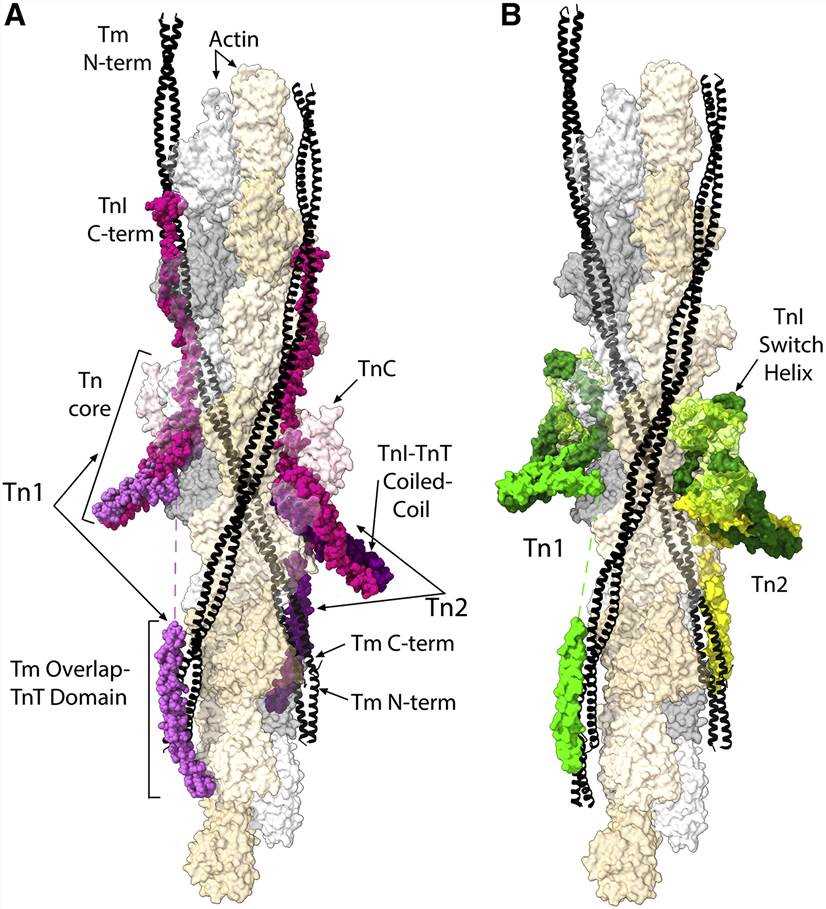 Figure 1. Atomic models of the cardiac thin filament in the apo-state and Ca2+-saturated state
(Source: Tobacman L S. 2021)
The Troponin T (TnT) subunit is the largest among the three troponin subunits and plays a crucial structural role. It anchors the troponin complex on the surface of the actin filaments and determines the orientation of the troponin core relative to the actin filaments. The subunit is structurally divided into a variety of isoforms that are differentially expressed in the heart, fast and slow muscle. In adult myocardium, the predominant isoform is the 35.9kDa TnT isoform T3. During protein processing, the N-terminal methionine is cleaved and consecutive serines are acetylated to produce a mature cardiac troponin T molecule with a length of 287 amino acids.
The amino acid sequence of troponin T is divided into three structural domains: the N-terminal domain, the linker domain, and the C-terminal domain. The C-terminal domain is part of the core troponin complex, while the remainder is associated with the pro-tropomyosin. The N-terminal fragment, called TnT1, has an extended structure that includes a single alpha helix region bound to the pro-tropomyosin, which may play a role in stabilizing the overlap region between the two pro-tropomyosin molecules.
 Figure 2. The cTnT molecule with its relevant structural regions highlighted
(Source: Marston S, et al. 2020)
cTnT has important clinical diagnostic and prognostic value, particularly in acute myocardial infarction (AMI) and in the assessment of cardiac injury. The elevated level of cTnT in blood indicates the damage of cardiomyocytes, which can be diagnosed and intervened early in heart-related diseases. Due to its high sensitivity and specificity, cTnT has been widely used in emergency rooms and intensive care units. Continuous monitoring of cTnT levels will help doctors understand the degree of myocardial injury and predict prognosis.
Troponin I (TnI) is a critical component of the troponin complex, primarily responsible for inhibiting muscle contraction. In a relaxed muscle state, TnI binds tightly to actin, preventing the interaction between actin and myosin, thus inhibiting muscle contraction. TnI is expressed in three isoforms: cardiac TnI (cTnI), slow skeletal muscle TnI, and fast skeletal muscle TnI. During embryonic development, slow skeletal muscle TnI is expressed in cardiac tissue, but it is replaced by cTnI after birth, which is then exclusively expressed in cardiac tissue. The mature form of human cTnI comprises a 210-residue protein (209 residues after removing the first methionine residue) with a molecular weight of approximately 24kDa. The protein structure includes four alpha helices and a flexible, disordered region in the middle. Structurally, it can be divided into the following domains: the N-terminal domain (NcTnI) unique to the heart, the structurally rigid IT arm, the "inhibitory" peptide, the switch peptide, and the C-terminal domain. Among them, the NcTnI region is only expressed in the cardiac TnI isoform; the IT arm is a rigid coiled coil of the α-helix of cTnI and cTnT; helix H2 is the "inhibitory peptide"; and helix H3 is the switch peptide. Like cTnT, cTnI is widely used in the clinical diagnosis of acute myocardial infarction and in the evaluation of cardiac injury. Elevated levels of cTnI in the blood indicate myocardial cell damage, which is crucial for early diagnosis and intervention in heart conditions.
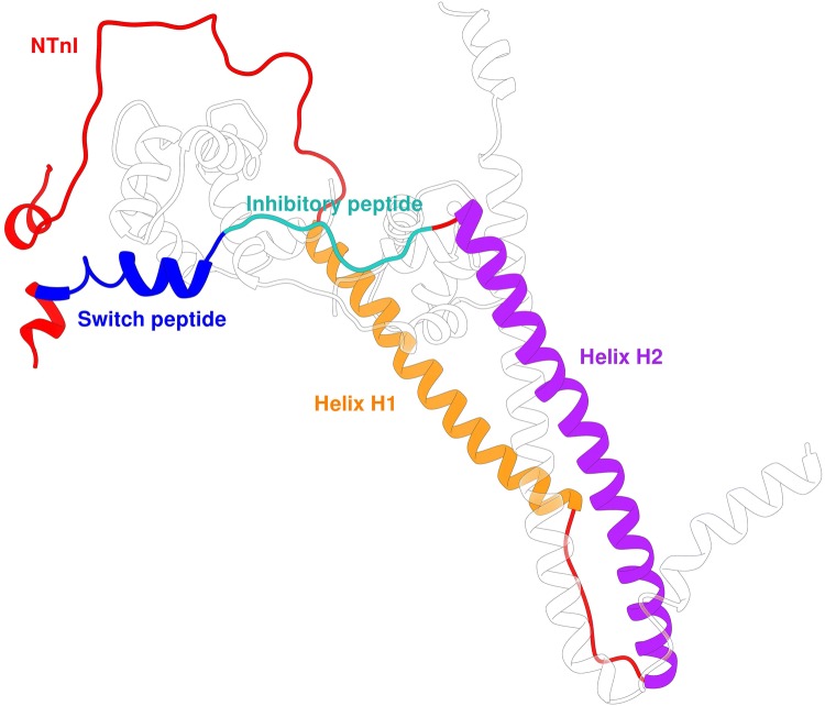 Figure 3. The cTnI molecule with its relevant structural regions highlighted
(Source: Marston S, et al. 2020)
As previously mentioned, the troponin test is a crucial diagnostic tool primarily used in cardiology to assess and monitor cardiac conditions. This test measures the levels of cardiac-specific troponins - cardiac troponin I and cardiac troponin T - in the blood. The most common assays currently in use are immunochemical methods, including enzyme-linked immunosorbent assay (ELISA), immunofluorescence assay, radioimmunoassay (RIA), and immunochemiluminescence assay. These assays share a similar foundational principle: in the immunological phase, a specific antibody (anti-cTn) binds to an antigen (cTn N-terminal amino acid). The secondary phase involves either an enzymatic reaction or an additional antibody-antigen interaction, depending on the method. Finally, the detection phase varies by assay type: a spectrophotometer is used to measure color intensity in ELISA, a radiometer detects radionuclide emissions in RIA, and a fluorometer detects the fluorophore in immunofluorescence. The intensity of the signal produced is proportional to the number of troponin molecules detected, allowing for the quantification of troponin levels. This precise measurement is instrumental in helping clinicians diagnose diseases and manage cardiac damage effectively.
The high-sensitivity troponin (hs-troponin) assay technology enables the detection of troponin at concentrations 10 to 100 times lower than conventional assays through improved antibody technology and signal detection methods, while ensuring, among other things, assay accuracy and reproducibility. Designed to detect very small amounts of cTnI and cTnT in the blood, the technology can identify myocardial damage much earlier than conventional tests, which is critical for timely intervention and treatment of AMI. In addition, elevated levels of hs-cTn provide an excellent prognostic indicator for patients with congenital heart disease, atrial fibrillation, heart failure and other conditions.
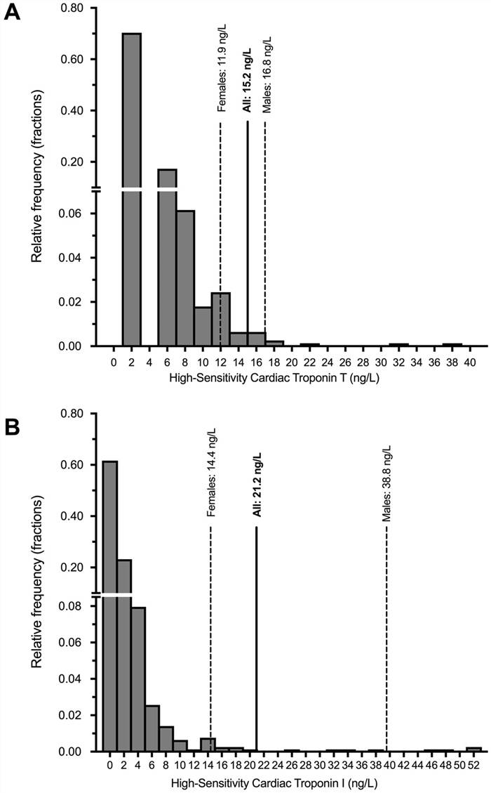 Figure 4. Distribution of high-sensitivity cardiac troponin T (Panel A) and I (Panel B) Concentrations in Cardio-renal Healthy Asians
(Source: Aw TC, et al. 2019)
References
| Target | Cat. No. | Product Name | Size | Species Reactivity | Application | Detection Sample | |
| Troponin T | DEIA-FN1574 | Rat Tn-T(Troponin T) ELISA Kit | 96T | Rat | Quantitative | Serum, plasma, cell culture supernatant and other biological samples | Inquiry |
| DEIA-FN1575 | Rabbit Tn-T (Troponin T) ELISA Kit | 96T | Quantitative | Serum, plasma, cell culture supernatants, tissue homogenate | Inquiry | ||
| DEIA-FN1575NS | Rabbit cTnT/TNNT2(Troponin T Type 2, Cardiac) ELISA Kit | 96T | Rabbit | Quantitative | Serum, plasma, cell culture supernatant and other biological samples | Inquiry | |
| Troponin I | DEIA-BJ2851 | Porcine Troponin Ⅰ ELISA Kit | 96T | Quantitative | Serum, plasma, cell culture supernatants, body fluid and tissue homogenate | Inquiry | |
| DEIA-BJ2307 | Rat Troponin Ⅰ ELISA Kit | 96T | Quantitative | Serum, plasma, cell culture supernatants, body fluid and tissue homogenate | Inquiry | ||
| TNNI3 | DEIA6156 | High Sensitivity Mouse Cardiac Troponin-I ELISA Kit | 96T | Mouse | Quantitative | Serum | Inquiry |
| DEIA-NS2307-89 | Monkey cTn-I/TNNI3(cardiac Troponin I) ELISA Kit | 96T | Monkey | Quantitative | Serum, plasma, cell culture supernatant and other biological samples. | Inquiry | |
| DEIA-NS2307-93 | Rat cTn-I/TNNI3(Cardiac Troponin I) ELISA Kit | 96T | Rat | Quantitative | Serum, plasma, tissue homogenates and other biological fluids. | Inquiry | |
| DEIA-BJ2846 | Porcine Cardiac Troponin 1 ELISA Kit | 96T | Quantitative | Serum, plasma, cell culture supernatants, body fluid and tissue homogenate | Inquiry | ||
| cTnI | DEIA-BJ2611 | Mouse CTNI(Cardiac Troponin-I) ELISA Kit | 96T | Mouse | Quantitative | Serum, plasma, tissue homogenates and other biological fluids | Inquiry |
| DEIACL19 | CDSimple™ cTnI Chemiluminescent ELISA Kit | 96T, 192T | Quantitative | Serum | Inquiry |
| Target | Cat. No. | Product Name | Expression System | Tag/Conjugate | Application | |
| Troponin T1 + T2 + T3 | DAGA-892 | Bovine troponin I-T-C complex | Bovine | Unconjugated | N/A | Inquiry |
| DAGA-880 | Cardiac troponin complex protein | Human heart tissue | Unconjugated | N/A | Inquiry | |
| DAGA-890 | Canine troponin I-T-C complex | Dog | Unconjugated | N/A | Inquiry | |
| cTnI | DAG-WT2214 | Recombinant Human cTnI [His] | E. coli | His | Immunoassays | Inquiry |
| TNNT1 | DAGA-909 | Bovine troponin I skeletal muscle (>98%) | Bovine | Unconjugated | N/A | Inquiry |
| DAGA-900 | Bovine troponin I cardiac (>98%) | Bovine | Unconjugated | N/A | Inquiry | |
| TNNT2 | DAGA-923 | Bovine troponin T skeletal muscle (>98%) | Bovine | Unconjugated | N/A | Inquiry |
| DAGA-916 | Bovine troponin T (>98%) | Bovine | Unconjugated | N/A | Inquiry | |
| TNNC1 | DAGA-882 | Recombinant Troponin C | E. coli | TBD | N/A | Inquiry |
| DAGA-329 | Recombinant Troponin I-Cardiac [His] | E. coli | His | ELISA | Inquiry |
| Target | Cat. No. | Product Name | Host | Isotype | Application | |
| Troponin T1 + T2 + T3 | DCABH-9396 | Anti-Troponin T1 + T2 + T3 monoclonal antibody, clone UU-09 (KMU-23) | Mouse | IgG1 | WB, IHC-P, IHC-Fr | Inquiry |
| Troponin T1 | DMABT-Z60249 | Anti-Drosophila melanogaster Troponin T1 Monoclonal antibody, Clone OCE-257 | Rat | IgG2a | ICC/IF, EM, WB | Inquiry |
| TNNC1 | DCABH-10062 | Anti-Cardiac Troponin monoclonal antibody, clone 31D7 | Mouse | IgG2b | ELISA, sELISA | Inquiry |
| TNNI3 | DPABH-27455 | Magic™ Anti-cardiac Troponin I (Phospho S105) polyclonal antibody | Rabbit | IgG | WB | Inquiry |
| TNNT2 | DPAB-DC4840 | Anti-Troponin I-Cardiac polyclonal antibody | Goat | IHC, ELISA, sELISA | Inquiry | |
| DPAB-DC4841 | Anti-Troponin I-Cardiac polyclonal antibody | Goat | IHC, ELISA, sELISA | Inquiry | ||
| TNNT1 | CABT-B1329 | Anti-Troponin T (Internal) monoclonal antibody, clone 5B5.2 | Mouse | IgG2a, κ | WB, IHC-P | Inquiry |
| TNNI1 | CABT-B275 | Anti-Troponin T, Striated Muscle monoclonal antibody, clone 53/36 | Mouse | IgG1 | WB | Inquiry |
Loading ......
